. 2009 Jul 29;422(1):23-35.
doi: 10.1042/BJ20090428.

Immunocytochemical techniques reveal multiple, distinct cellular pools of PtdIns4P and PtdIns(4,5)P(2)

Affiliations

Immunocytochemical techniques reveal multiple, distinct cellular pools of PtdIns4P and PtdIns(4,5)P(2)

Gerald R V Hammond et al. Biochem J. .

Abstract

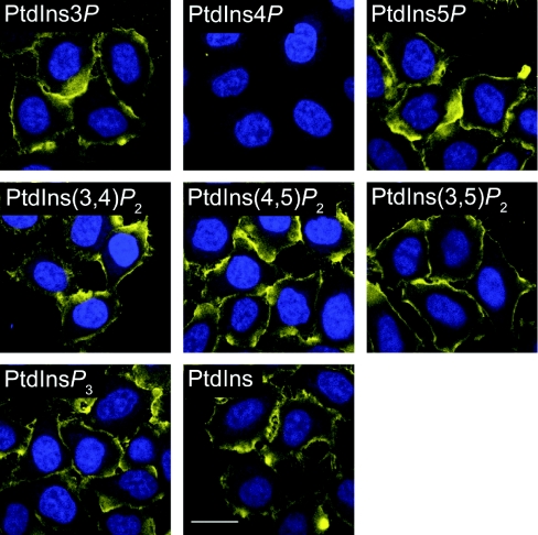
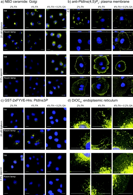
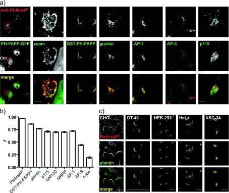
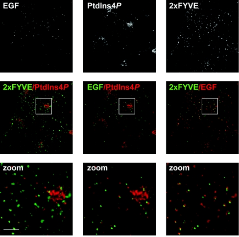
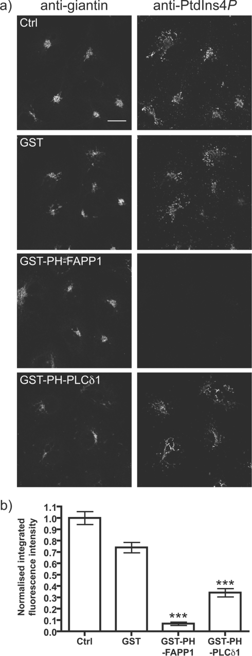
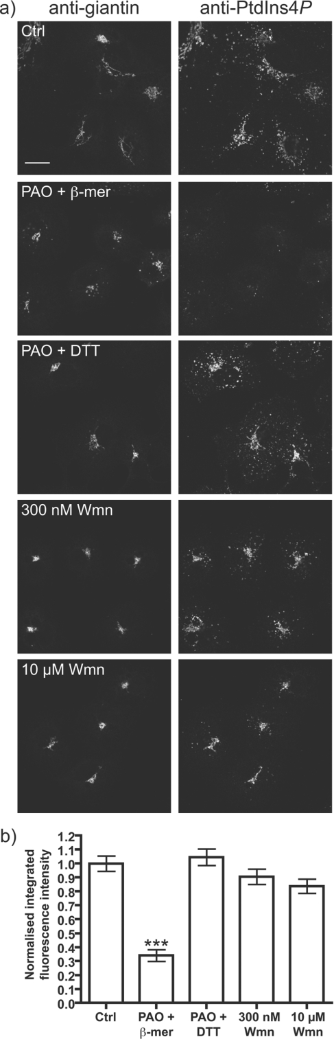
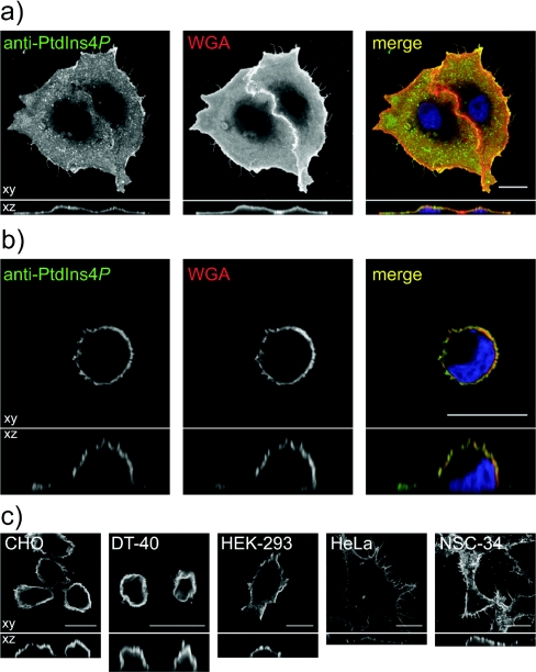
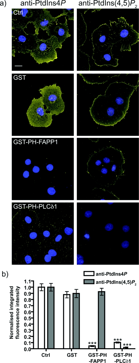
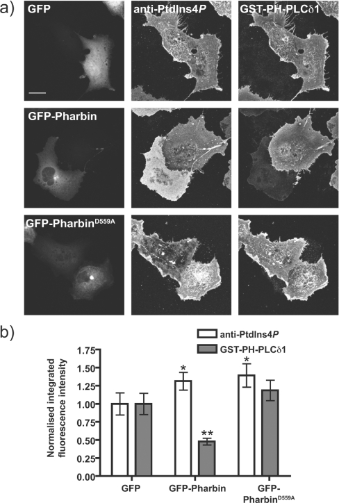
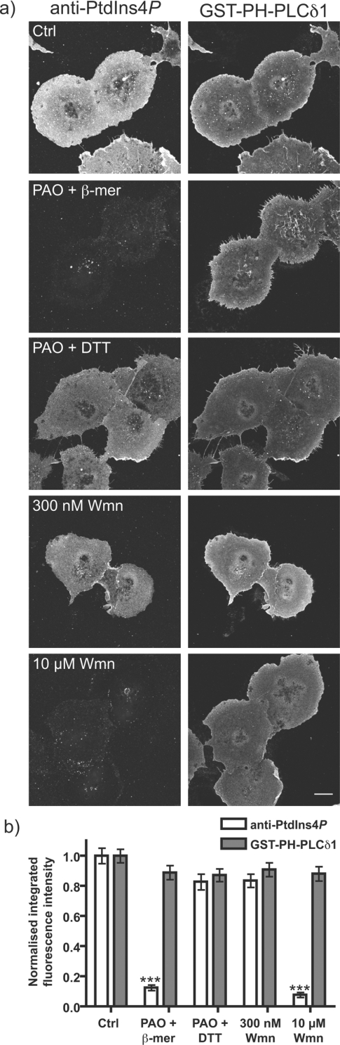
PtdIns4P is the major precursor for the synthesis of the multifunctional plasma membrane lipid, PtdIns(4,5)P(2). Yet PtdIns4P also functions as a regulatory lipid in its own right, particularly at the Golgi apparatus. In the present study we define specific conditions that enable preservation of several organellar membranes for the immunocytochemical detection of PtdIns4P. We report distinct pools of this lipid in both Golgi and plasma membranes, which are synthesized by different PI4K (phosphatidylinositol 4-kinase) activities, and also the presence of PtdIns4P in cytoplasmic vesicles, which are not readily identifiable as PI4K containing trafficking intermediates. In addition, we present evidence that the majority of PtdIns4P resides in the plasma membrane, where it is metabolically distinct from the steady-state plasma membrane pool of PtdIns(4,5)P(2).


Figures

Figure 1
Figure 1. Specificity of the anti-PtdIns4P antibody
HeLa cells were fixed with 4% FA plus 0.2% GA and stained on ice using saponin permeabilization, as described in the Experimental section. Anti-PtdIns4P antibody was pre-absorbed with POPC liposomes containing 5% (mol/mol) of the indicated inositol lipid for 1 h at room temperature prior to applying to the cells on ice. Anti-PtdIns4P antibody staining is yellow, and DAPI-stained nuclei are in blue. Scale bar=20 μm. Images are single confocal optical sections.
Figure 2
Figure 2. Effect of fixation, detergent and temperature on the retention of various organelle-specific lipid stains
Cells were fixed with the indicated concentration of aldehydes, then stained at the designated temperature after permeabilization with digitonin or saponin. See the Experimental section for details. Agents were used that specifically label the (a) Golgi (NBD-ceramide), (b) plasma membrane (anti-PtdIns(4,5)P2 antibody), (c) endosomes (anti-GST against GST–2×FYVE-Hrs bound to PtdIns3P) or (d) endoplasmic reticulum (DiOC6). DAPI-stained nuclei are shown in blue. Scale bars=20 μm.
Figure 3
Figure 3. Anti-PtdIns4P antibody labels the Golgi apparatus at room temperature after 2% FA fixation and digitonin permeabilization
(a) COS-7 cells were stained with anti-PtdIns4P antibody. Where indicated cells were expressing a GFP-tagged PH domain from FAPP1 (expressed for 24 h) and were co-stained using either an anti-GST antibody (against the purified GST-tagged FAPP1 PH domain) or antibodies against the indicated markers. Scale bars=5 μm. (b) Pearson's correlation coefficients for co-localized intensity were calculated between labelling for PtdIns4P and the indicated marker, as described in the Experimental section. Images not shown in (a) are in Supplementary Figure S2 at http://www.BiochemJ.org/bj/422/bj4220023add.htm. Results are presented as means±S.E.M. and a statistical comparison is shown in Table 1. (c) PtdIns4P and giantin labelling in a variety of cell types. Scale bars=20 μm. Images are single confocal optical sections.
Figure 4
Figure 4. Co-localization of PtdIns4P and PtdIns3P with internalized EGF
COS-7 cells were serum-starved overnight, then incubated for 30 min with 10 μg/ml Alexa Fluor® 488-conjugated EGF, prior to fixing and staining for PtdIns4P and PtdIns3P as described in Figure 3. Images labelled ‘zoom’ show the enlarged region outlined in the panels above. Scale bars=5 μm. Images are single confocal optical sections.
Figure 5
Figure 5. Competition for Golgi PtdIns4P staining by PH-FAPP1
(a) The indicated GST-tagged fusion proteins (10 μM) were included with the primary anti-PtdIns4P antibody and anti-giantin antibodies applied to COS-7 cells. Staining conditions are as Figure 3. Single confocal optical sections are shown. Scale bar=20 μm. (b) Quantification of anti-PtdIns4P antibody labelling (n=207). See the Experimental section for details. ***P<0.001.
Figure 6
Figure 6. Effect of PI4K inhibitors on Golgi PtdIns4P labelling
(a) COS-7 cells were pre-treated for 30 min at 37 °C with 0.1% DMSO (Ctrl), 10 μM PAO with 1 mM 2-mercaptoethanol or DTT (PAO+β-mer or PAO+DTT) and 300 nM or 10 μM wortmannin (Wmn) as indicated, prior to fixation and staining as described in Figure 3. Single confocal optical sections are shown. Scale bar=20 μm. (b) Quantification of anti-PtdIns4P labelling (n=237). See Experimental section for details. *** P<0.001.
Figure 7
Figure 7. Anti-PtdIns4P antibody labels the plasma membrane of cells fixed with FA and GA, permeabilized with saponin and stained on ice
(a) COS-7 cells grown overnight prior to fixation and staining or (b) fixed and stained 20 min after seeding on poly-L-lysine. In both cases, cells were incubated for 5 min on ice with 50 μg/ml Alexa Fluor® 555-conjugated WGA prior to fixing to label the plasma membrane. (c) Different cell types stained with anti-PtdIns4P antibody using the above conditions. All images show single confocal optical sections in the xy plane (upper panels), or sections through the z plane (lower panels). Scale bars=20 μm.
Figure 8
Figure 8. Competition for plasma membrane PtdIns4P staining by PH-FAPP1
(a) The indicated GST-tagged fusion proteins were included at 10 μM with the primary anti-PtdIns4P or anti-PtdIns(4,5)P2 antibodies applied to COS-7 cells. Staining conditions are as in Figure 7. Images are maximum intensity projections of confocal sections spanning the entire depth of the cells. Scale bar=20 μm. DAPI stained nuclei are in blue indicating the presence of cells where lipid staining is absent. (b) Quantification of staining (n=126 cells for anti-PtdIns4P antibody and 123 cells for anti-PtdIns(4,5)P2 antibody). See the Experimental section for details. ***P<0.001.
Figure 9
Figure 9. Effect of a 5-phosphatase on plasma membrane PtdIns4P/PtdIns(4,5)P2 labelling
COS-7 cells were transfected with GFP, GFP-Pharbin or the putatively inactive mutant of the latter, D559A, for 18 h prior to fixation and staining as described in Figure 7. (a) Representative images of COS-7 cells transfected with each construct. Images are maximum intensity projections of confocal sections spanning the entire depth of the cells. Scale bar=20 μm. (b) Quantification of staining (n=93 cells). See the Experimental section for details. *P<0.05, **P<0.01.
Figure 10
Figure 10. Effect of PI4K inhibitors on plasma membrane PtdIns4P and PtdIns(4,5)P2 labelling
(a) COS-7 cells were pre-treated for 30 min at 37 °C with 0.1% DMSO (Ctrl), 10 μM PAO with 1 mM 2-mercaptoethanol or DTT (PAO+β-mer or PAO+DTT), 300 nM or 10 μM wortmannin (Wmn) as indicated, prior to fixation and staining with anti-PtdIns4P antibody and GST–PH-PLCδ1 as described in Figure 7. Images are maximum intensity projections of confocal sections spanning the entire depth of the cells. Scale bar=20 μm. (b) Quantification of staining (n=136 cells). See the Experimental section for details. ***P<0.001.

References

    1. Balla T. Phosphoinositide-derived messengers in endocrine signaling. J. Endocrinol. 2006;188:135–153. - PubMed
    1. Vanhaesebroeck B., Leevers S. J., Ahmadi K., Timms J., Katso R., Driscoll P. C., Woscholski R., Parker P. J., Waterfield M. D. Synthesis and function of 3-phosphorylated inositol lipids. Annu. Rev. Biochem. 2001;70:535–602. - PubMed
    1. Balla A., Balla T. Phosphatidylinositol 4-kinases: old enzymes with emerging functions. Trends Cell Biol. 2006;16:351–361. - PubMed
    1. Di Paolo G., De Camilli P. Phosphoinositides in cell regulation and membrane dynamics. Nature. 2006;443:651–657. - PubMed
    1. D'angelo G., Vicinanza M., Di Campli A., De Matteis M. The multiple roles of PtdIns(4)P – not just the precursor of PtdIns(4,5)P2. J. Cell Sci. 2008;121:1955–1963. - PubMed

Publication types

MeSH terms