Comparative morphology of the axial complex and interdependence of internal organ systems in sea urchins (Echinodermata: Echinoidea)
- PMID: 19508706
- PMCID: PMC2701938
- DOI: 10.1186/1742-9994-6-10
Comparative morphology of the axial complex and interdependence of internal organ systems in sea urchins (Echinodermata: Echinoidea)
Abstract
Background: The axial complex of echinoderms (Echinodermata) is composed of various primary and secondary body cavities that interact with each other. In sea urchins (Echinoidea), structural differences of the axial complex in "regular" and irregular species have been observed, but the reasons underlying these differences are not fully understood. In addition, a better knowledge of axial complex diversity could not only be useful for phylogenetic inferences, but improve also an understanding of the function of this enigmatic structure.
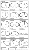
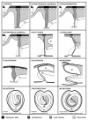
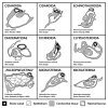
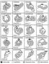
Results: We therefore analyzed numerous species of almost all sea urchin orders by magnetic resonance imaging, dissection, histology, and transmission electron microscopy and compared the results with findings from published studies spanning almost two centuries. These combined analyses demonstrate that the axial complex is present in all sea urchin orders and has remained structurally conserved for a long time, at least in the "regular" species. Within the Irregularia, a considerable morphological variation of the axial complex can be observed with gradual changes in topography, size, and internal architecture. These modifications are related to the growing size of the gastric caecum as well as to the rearrangement of the morphology of the digestive tract as a whole.
Conclusion: The structurally most divergent axial complex can be observed in the highly derived Atelostomata in which the reorganization of the digestive tract is most pronounced. Our findings demonstrate a structural interdependence of various internal organs, including digestive tract, mesenteries, and the axial complex.
Figures


Similar articles
-
Origin and evolutionary plasticity of the gastric caecum in sea urchins (Echinodermata: Echinoidea).BMC Evol Biol. 2010 Oct 18;10:313. doi: 10.1186/1471-2148-10-313. BMC Evol Biol. 2010. PMID: 20955602 Free PMC article.
-
Systematic comparison and reconstruction of sea urchin (Echinoidea) internal anatomy: a novel approach using magnetic resonance imaging.BMC Biol. 2008 Jul 23;6:33. doi: 10.1186/1741-7007-6-33. BMC Biol. 2008. PMID: 18651948 Free PMC article.
-
A dataset comprising 141 magnetic resonance imaging scans of 98 extant sea urchin species.Gigascience. 2014 Oct 14;3:21. doi: 10.1186/2047-217X-3-21. eCollection 2014. Gigascience. 2014. PMID: 25356198 Free PMC article.
-
Echinoderms: their culture and bioactive compounds.Prog Mol Subcell Biol. 2005;39:139-65. Prog Mol Subcell Biol. 2005. PMID: 17152697 Review.
-
Arrays in rays: terminal addition in echinoderms and its correlation with gene expression.Evol Dev. 2005 Nov-Dec;7(6):542-55. doi: 10.1111/j.1525-142X.2005.05058.x. Evol Dev. 2005. PMID: 16336408 Review.
Cited by
-
Evolution of a novel muscle design in sea urchins (Echinodermata: Echinoidea).PLoS One. 2012;7(5):e37520. doi: 10.1371/journal.pone.0037520. Epub 2012 May 18. PLoS One. 2012. PMID: 22624043 Free PMC article.
-
Inside out: modern imaging techniques to reveal animal anatomy.PLoS One. 2011 Mar 22;6(3):e17879. doi: 10.1371/journal.pone.0017879. PLoS One. 2011. PMID: 21445356 Free PMC article.
-
Applications of chemical shift imaging to marine sciences.Mar Drugs. 2010 Aug 19;8(8):2369-83. doi: 10.3390/md8082369. Mar Drugs. 2010. PMID: 20948912 Free PMC article.
-
Origin and evolutionary plasticity of the gastric caecum in sea urchins (Echinodermata: Echinoidea).BMC Evol Biol. 2010 Oct 18;10:313. doi: 10.1186/1471-2148-10-313. BMC Evol Biol. 2010. PMID: 20955602 Free PMC article.
-
Magnetic resonance imaging (MRI) has failed to distinguish between smaller gut regions and larger haemal sinuses in sea urchins (Echinodermata: Echinoidea).BMC Biol. 2009 Jul 13;7:39; author reply 39. doi: 10.1186/1741-7007-7-39. BMC Biol. 2009. PMID: 19594924 Free PMC article.
References
-
- Ruppert EE. Introduction to the aschelminth phyla: a consideration of mesoderm, body cavities, and cuticle. In: Harrison FW, Ruppert EE, editor. Microscopic Anatomy of Invertebrates. Vol. 4. New York: Wiley-Liss; 1991. pp. 1–17.
-
- Bartolomaeus T. On the ultrastructure of the coelomic lining in annelids, echiurids and sipunculids. Microfauna Marina. 1994;9:171–220.
-
- Rieger RM, Purschke G. The coelom and the origin of the annelid body plan. Hydrobiologia. 2005;535:127–137. doi: 10.1007/s10750-004-4381-6. - DOI
-
- Heider K. Über Organverlagerungen bei der Echinodermen-Metamorphose. Verhandlungen der Deutschen Zoologischen Gesellschaft. 1912;22:239–251.
-
- Ruppert EE, Balser EJ. Nephridia in the larvae of hemichordates and echinoderms. Biological Bulletin. 1986;171:188–196. doi: 10.2307/1541916. - DOI
LinkOut - more resources
Full Text Sources
Miscellaneous

