Review
. 2009 Oct;20(8):978-85.
doi: 10.1016/j.semcdb.2009.02.007.

Line up and listen: Planar cell polarity regulation in the mammalian inner ear

Affiliations
Review

Line up and listen: Planar cell polarity regulation in the mammalian inner ear

Padmashree C G Rida et al. Semin Cell Dev Biol. 2009 Oct.

Abstract

The inner ear sensory organs possess extraordinary structural features necessary to conduct mechanosensory transduction for hearing and balance. Their structural beauty has fascinated scientists since the dawn of modern science and ensured a rigorous pursuit of the understanding of mechanotransduction. Sensory cells of the inner ear display unique structural features that underlie their mechanosensitivity and resolution, and represent perhaps the most distinctive form of a type of cellular polarity, known as planar cell polarity (PCP). Until recently, however, it was not known how the precise PCP of the inner ear sensory organs was achieved during development. Here, we review the PCP of the inner ear and recent advances in the quest for an understanding of its formation.


Figures
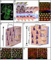
Fig. 1
Fig. 1. Planar cell polarity in the sensory epithelia of the inner ear
(A) The inner ear consists of 6 sensory epithelia marked by the expression of GFP: the organ of Corti of the cochlea (CO), the maculae of the saccule (SA) and utricle (UT), and the three orthogonally positioned cristae: the anterior crista (AC), posterior crista (PC) and lateral crista (LC). The white tracing outlines the fluid-filled labyrinth of the inner ear. (B-G) Schematic diagrams (B, D, F) and confocal images (C, E, G) illustrating the planar polarity of the organ of Corti (B-C), LC (D-E), and Ut (F-G). In the organ of Corti (B), the inner (IHCs) and outer hair cells (OHCs) are interdigitated with several distinct types of nonsensory supporting cells: the inner phalangeal cells (IPHs), inner pillar cells (IPCs), outer pillar cells (OPCs) and three rows of Deiters’ cells (DC1-DC3s). The kinocilia (BC, blue) and stereocilia (B, purple, C, green) constitute a “V”-shaped structure and all the “V”s are uniformly aligned, showing a distinctive PCP. In LC (E), Spectrin (red) accumulates in the cuticular plates of the hair cells but is conspicuously excluded from the fonticulus within which the basal body resides, providing a convenient marker for hair cell polarity. The hair bundles and cell membranes are visualized by staining for actin (green). (G) In Ut, Spectrin staining (red) permits easy visualization of the intrinsic polarity of each hair cell (white arrows) while cell membranes can be seen by staining for actin (green). The hair cells on the two sides of the line of polarity reversal (pink line) are oriented towards each other.
Fig2
Fig2. Distinct cellular mechanisms underlying the cochlear phenotypes in core PCP and ciliary mutants
(A) The cochlea in a PCP mutant (A) or a ciliary mutant (not shown) embryo is markedly shorter and wider than that of wild-type embryo, indicating that both core PCP and ciliary genes are required for CE of the cochlea. (B-D) Compared to wild-type (B), core PCP (C) and ciliary (D) mutants share hair bundle misorientation defects (asterisks). In ciliary mutants (D), circular hair bundles were also present (white arrowheads), indicating the loss of the intrinsic polarity of hair cells that is unique to ciliary mutants. (E-I) Polarized distribution of core PCP proteins in wild-type (E, F) is lost in PCP mutant (G), but retained in ciliary mutants, even when hair bundles have rotated away from their normal orientation (H, I). (I) is a close-up of the cell outlined by a dashed square in the panel (H).M: medial; L: lateral
Fig. 3
Fig. 3. Model showing the formation of planar cell polarity in the cochlea
PCP establishment in the cochlea involves at least 3 main morphogenetic stages: (A) Kinocilia initially protrude from the center of the apical surface of the newly post-mitotic cells in the sensory primordium of the cochlea. (B) Polarization of core PCP proteins, including Vangl2, Fz3 and Fz6, and Dvl2 and Dvl3, and eccentric localization of primary cilia and centriole alignment along the PCP axis. The interaction between Fz and Vang is hypothesized. Other core PCP proteins, such as Pk and Dvl, may preferentially interact with Fz or Vang to reinforce polarized sorting of Vang and Fz within the same cell and to recruit downstream factors for localized cytoskeletal remodeling. (C) Formation of uniformly oriented hair bundles in the organ of Corti. Both core PCP genes and ciliary genes are clearly required for morphological polarity of the organ of Corti. Series of red arrows in (B) indicate potential effectors downstream of core PCP proteins that target at actin and microtubule cytoskeletons to achieve morphological polarity. Asterisk indicates genes that have been shown to repress canonical Wnt signaling. They include Wnt coreceptor Ror, Diversin/Ankrd6, and ciliary and basal body genes. M: medial (center of the cochlear spiral), L: lateral (periphery of the cochlear spiral).

References

    1. Gubb D, Garcia-Bellido A. A genetic analysis of the determination of cuticular polarity during development in Drosophila melanogaster. J Embryol Exp Morphol. 1982;68:37–57. - PubMed
    1. Keller RE, Danilchik M, Gimlich R, Shih J. The function and mechanism of convergent extension during gastrulation of Xenopus laevis. J Embryol Exp Morphol. 1985;89(Suppl):185–209. - PubMed
    1. Keller R, Tibbetts P. Mediolateral cell intercalation in the dorsal, axial mesoderm of Xenopus laevis. Dev Biol. 1989;131:539–49. - PubMed
    1. Wallingford JB, Rowning BA, Vogeli KM, Rothbacher U, Fraser SE, Harland RM. Dishevelled controls cell polarity during Xenopus gastrulation. Nature. 2000;405:81–5. - PubMed
    1. Klein TJ, Mlodzik M. Planar cell polarization: an emerging model points in the right direction. Annu Rev Cell Dev Biol. 2005;21:155–76. - PubMed

Publication types

LinkOut - more resources