. 2009 Aug;58(8):1797-806.
doi: 10.2337/db09-0317. Epub 2009 Jun 9.

Mesenchymal stem cells prevent the rejection of fully allogenic islet grafts by the immunosuppressive activity of matrix metalloproteinase-2 and -9

Affiliations

Mesenchymal stem cells prevent the rejection of fully allogenic islet grafts by the immunosuppressive activity of matrix metalloproteinase-2 and -9

Yunchuan Ding et al. Diabetes. 2009 Aug.

Abstract

Objective: Mesenchymal stem cells (MSCs) are known to be capable of suppressing immune responses, but the molecular mechanisms involved and the therapeutic potential of MSCs remain to be clarified.

Research design and methods: We investigated the molecular mechanisms underlying the immunosuppressive effects of MSCs in vitro and in vivo.

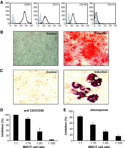
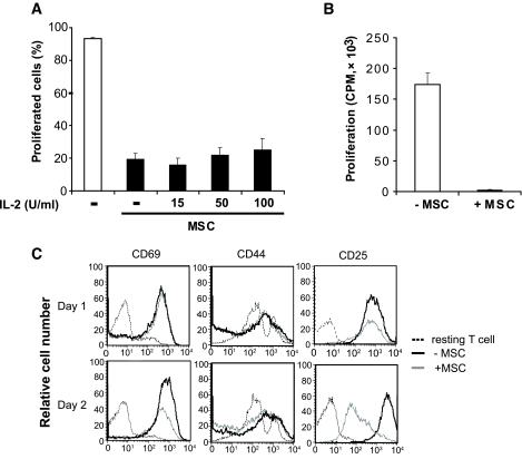
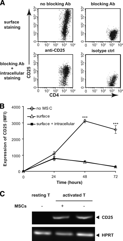
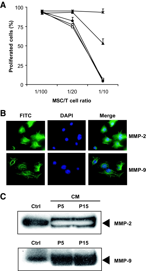
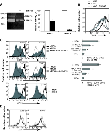
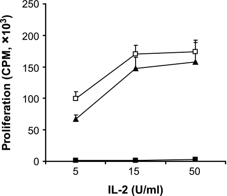
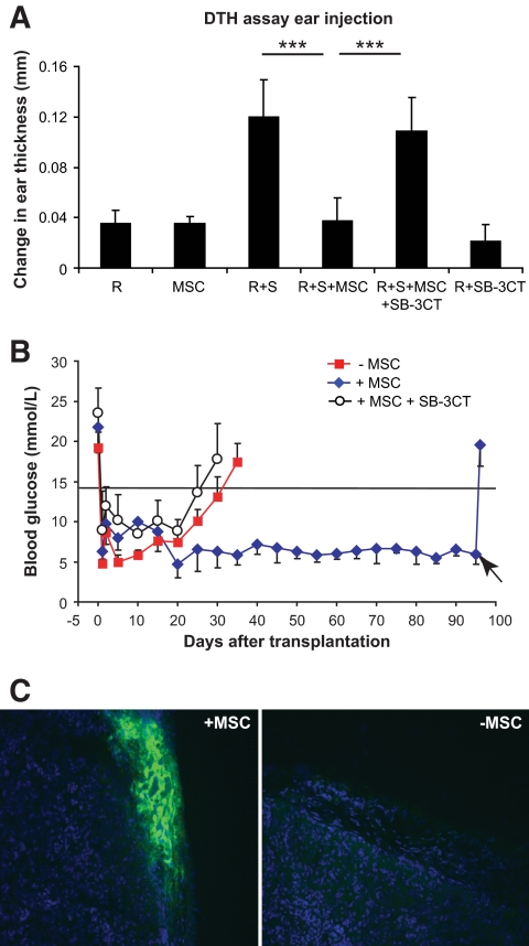
Results: Our results demonstrate that matrix metalloproteinases (MMPs) secreted by MSCs, in particular MMP-2 and MMP-9, play an important role in the suppressive activity of MSCs by reducing surface expression of CD25 on responding T-cells. Blocking the activity of MMP-2 and MMP-9 in vitro completely abolished the suppression of T-cell proliferation by MSCs and restored T-cell expression of CD25 as well as responsiveness to interleukin-2. In vivo, administration of MSCs significantly reduced delayed-type hypersensitivity responses to allogeneic antigen and profoundly prolonged the survival of fully allogeneic islet grafts in transplant recipients. Significantly, these MSC-mediated protective effects were completely reversed by in vivo inhibition of MMP-2 and MMP-9.

Conclusions: We demonstrate that MSCs can prevent islet allograft rejection leading to stable, long-term normoglycemia. In addition, we provide a novel insight into the mechanism underlying the suppressive effects of MSCs on T-cell responses to alloantigen.


Figures

FIG. 1.
FIG. 1.
MSCs suppress T-cell proliferation in response to anti-CD3/CD28 cross linking or allogeneic antigen stimulation. A: MSCs obtained from bone marrow cells of BALB/c (H2d) mice show spindle-shaped morphology and exhibit a typical MSC phenotype (solid line), in which they are positive for CD44, CD73, and CD106 but negative for CD11b. Isotype controls are represented as the shaded curve. B: MSCs were able to differentiate into osteogenic and adipogenic lineages when subjected to inductive stimuli (B and C, induction) but not in control medium (B and C, control). Osteogenic differentiation of MSCs was verified by Alizarin red staining (B). The presence of intracellular fat droplets detected by Oil Red O staining indicates induced adipogenesis (C). D: CD4+CD25 T-cells were purified from C57BL/6 (H2b) mice using CD25 MicroBeads. Standard 3-day cultures were set up in 96-well round-bottomed plates using 2 × 10⁵ CFSE-labeled responder T-cells stimulated by CD3/CD28 Dynalbeads at a 1:1 bead:cell ratio in the presence of graded numbers of MSCs. CFSE division history was analyzed by flow cytometry. The percentage of proliferated cells was calculated by dividing the number of cells undergoing divisions with the total T-cells in the cultures. E: For mixed leukocyte cultures, 2 × 10⁵ CFSE-labeled CD4+CD25 T-cells were cocultured with 5 × 10⁵ irradiated (3,600 rad) CBA.Ca (H2k) splenocytes for 5 days. All the data above were obtained from three independent experiments. A high-quality digital representation of this figure is available in the online issue.
FIG. 2.
FIG. 2.
T-cell blasts cocultured with MSCs exhibited hyporesponsiveness to exogenous IL-2 and lost surface expression of CD25. A: CFSE-labeled CD4+CD25 T-cells from naïve C57BL/6 mice (H2b) were cultured at a density of 2 × 10⁵ per well in 96-well plates and stimulated with anti-CD3/CD28 beads alone or in the presence of MSCs (2 × 10⁴). Exogenous IL-2 was added at concentrations of 15, 50, and 100 IU/ml. After 72 h, cell proliferation was analyzed by flow cytometry. Data are represented as means ± SD. Results are the average of three experiments of identical design. B: Bars show the SD CD4+CD25 T-cells were stimulated with anti-CD3/CD28 beads alone or in the presence of MSCs (MSC:T-cell ratio of 1:10) for 72 h. CD4+ T-cells were purified by MACs anti-CD4 beads and cultured with 50 IU/ml IL-2 for 72 h. T-cell proliferation was measured by 3H-Tdr incorporation added during the last 8 h of culture. C: T-cells stimulated with anti-CD3/CD28 beads in the presence or absence of MSC and harvested after 24 and 48 h. Surface expression of CD69, CD44, and CD25 was evaluated by flow cytometry. Histograms are representative of three independent experiments.
FIG. 3.
FIG. 3.
Reduced expression of surface CD25 in the presence of MSCs is not related to receptor internalization or reduced transcriptional activity. A: As a positive control for intracellular CD25 detection, activated T-cells were incubated with unconjugated PC61 then stained with PC61-Pe with or without permeabilization or with an isotype control. B: CD4+CD25 T-cells from naïve C57BL/6 mice were stimulated with anti-CD3/CD28 beads and cocultured with MSCs for 24, 48, and 72 h followed by extracellular staining or extracellular plus intracellular staining for CD25. The surface expression of CD25 (MFI) was significantly reduced at 48 and 72 h in the presence of MSCs, which was not explained by an increase in intracellular CD25, since the surface and surface plus intracellular profiles are essentially identical. C: T-cells were stimulated with anti-CD3/CD28 beads in the presence or absence of MSCs at the MSC:T-cell ratio of 1:10. Total mRNA was extracted from bead-purified T-cells after coculture or from naïve T-cells, and cDNA was synthesized using CMV reverse transcriptase. For PCR, CD25 primers were as follows: CD25 forward primer 5′-GAA GCC AAC ACA GTC TAT GCA CC-3′ and CD25 reverse primer 5′-TGT CCT TCC ACG AAA TGA TAG ATT C-3′; and HPRT forward primer 5′-ATC ATT ATG CCG AGG ATT TGG AA-3′ and HPRT reverse primer 5′-TTG AGC ACA CAG AGG GCC A-3′.
FIG. 4.
FIG. 4.
MMP-2 and MMP-9 are involved in the immunosuppression by MSCs. A: CFSE-labeled CD4+CD25 T-cells stimulated with anti-CD3/CD28 were cultured for 72 h in the presence or absence of MSCs. Cell cultures were treated with 1 mmol/l 1-MT or 1 mmol/l l-NMM or 1 mmol/l SnPP or 6 μmol/l SB-3CT. Proliferation was accessed by dilution of CFSE fluorescent intensity. Data from three independent experiments each performed in triplicate are represented as means ± SD. □, control; ○, 1-MT; ▴, l-NMMA; ●, SnPP; *SB-3CT. B: MSCs were seeded on 13-mm glass coverslips and fixed, permeabilized, and blocked with serum and incubated with mouse anti–MMP-9 or anti–MMP-2 monoclonal antibodies. Binding was visualized using an Alexa Fluro 488 FITC-conjugated secondary, and cells were counterstained with Hoechst 33342. Sections were examined using a Leica epifluorescence microscope. C: To collect conditioned medium, MSCs were washed with PBS three times and resupplied with serum-free medium for 24 h. After harvest, the MSC-conditioned medium was spun at 1,500g to remove cellular debris, loaded on SDS-PAGE gels, and blotted with monoclonal anti–MMP-2 or –MMP-9 antibodies. Recombinant mouse MMP-2 and MMP-9 were used as markers. Data shown are representative of three independent experiments. A high-quality digital representation of this figure is available in the online issue.
FIG. 5.
FIG. 5.
MSC-dependent reduction in CD25 expression by T-cells is mediated by MMP-2 and MMP-9. A: Enzymatic activities of MMP-2 and MMP-9 in medium conditioned by MSCs were analyzed by zymography. Conditioned medium was electrophoresed under nonreducing conditions and without heating through a 8% SDS-PAGE containing 0.1% gelatin. Following electrophoresis, the gels were incubated overnight in developing buffer containing SB-3CT to inhibit MMP activity. Gels were stained with Coomassie Blue, and densitometric quantification of MMP-2 and MMP-9 was performed using Scion software. Data were obtained from three individual experiments, and inhibition of MMP-2 and MMP-9 was determined by setting the mock treatment as 100%. □, control; ■, SB-3CT. B: To block activities of MMP-2 and MMP-9 in the MSC/T-cell coculture, SB-3CT was added at a concentration of 6 μmol/l. Stimulated T-cells alone (black curve) or cocultured with MSCs (dashed curve) or MSCs treated with SB-3CT (gray curve) were examined for surface expression of CD25. C: Stimulated T-cells were either cultured alone (black curve) or in the presence of MSCs (MSC/T-cell ratio of 1:10) (dashed curve). In some experiments, neutralizing monoclonal anti–MMP-2 or –MMP-9 antibodies were added (gray shaded). Neutralizing antibody was used to block the activity of MT1-MMP. IgG1 was used as an isotype control. T-cells were harvested at 72 h, stained with CD25-PE, and analyzed by flow cytometry. D: CD4+CD25 T-cells were activated by 10 μg/ml ConA for 24 h, washed three times to remove residual ConA, and resuspended in serum-free medium at final concentration of 2 × 10⁵/ml. MMP-2 and MMP-9 recombinant proteins were added at final concentration of 1 mg/ml. T-cells were incubated with MMP recombinant proteins for an additional 6 h. MMP-2 or MMP-9 treated cells (solid curve) and untreated cells (dotted curve) were analyzed for CD25 expression by flow cytometry. Histograms of CD25 expression on T-cells are representative of three independent experiments.
FIG. 6.
FIG. 6.
Inhibition of MMP-2 and -9 reverses MSC-induced T-cell nonresponsiveness to IL-2. Anti-CD3/CD28–stimulated T-cells were cultured alone or in the presence of untreated MSCs or MSCs treated with SB-3CT. After 72 h, CD4+ T-cells were purified using MACs anti-CD4 beads and incubated with IL-2 at concentrations of 5, 15, and 50 IU/ml. Proliferation was measured by 3H-Tdr incorporation. Data from three independent experiments, each performed in triplicates, are presented as means ± SD. □, −MSC; ■, +MSC; ▴, +MSC + SB-3CT.
FIG. 7.
FIG. 7.
Prevention of DTH responses and protection of allogeneic islet grafts by MSCs is dependent on MMP-2 and MMP-9. A: 7 × 10⁶ responder cells (C57BL/6) or 7 × 10⁶ irradiated stimulator cells (CBA.Ca) were injected into the ear pinnae of BALB/c Rag−.−γ−.− mice either alone or together in the presence or absence of 1 ×10⁵ MSCs. DTH response–induced ear swelling was calculated by subtracting the thickness before injection from the thickness after 48 h. Administration of MSCs led to profound reduction of the alloreactive DTH response, which was reversed in the presence of the inhibitor SB-3CT. Data shown are means ± SD of a representative of at least three independent experiments. B: BALB/c Rag−.−γ−.− were rendered diabetic by a single intravenous injection of 200 mg/kg STZ. Mice had an average blood glucose concentration of 20 mmol/l immediately prior islet transplantation. A total of 500 IE pancreatic islets from CB57/B6 mice (H2b) alone or in the presence of 4 × 10⁵ BALB/c MSCs (H2d) were transplanted under the kidney capsules of STZ-induced diabetic BALB/c Rag−.−γ−.−. In some experiments, MSCs were pretreated with 6 μmol/l SB-3CT for 48 h and mice receiving treated MSCs also received SB-3CT (25 μg/mouse) intraperitoneally once every 4 days from the day of islet transplant until rejection. All islet transplant recipients were reconstituted with 1 × 10⁵ naïve BALB/c CD4+CD25 T-cells as an effector T-cell population. Rejection was defined as a blood glucose >14.5 mmol/l for at least 2 consecutive days. Continued function of the islet allografts was confirmed by removal of the islet-bearing kidneys and a return to hyperglycemia. C: Kidneys transplanted with islets grafts were excised at the time of rejection or the end point of this study. Immunofluorescence examination of islet grafts in MSC-treated recipients revealed intensely staining insulin producing islets beneath the kidney capsule, whereas in the MSC-untreated recipients the insulin-positive islet tissue was not detected at the time of rejection. A high-quality digital representation of this figure is available in the online issue.

Comment in

References

    1. Pittenger MF, Mackay AM, Beck SC, Jaiswal RK, Douglas R, Mosca JD, Moorman MA, Simonetti DW, Craig S, Marshak DR: Multilineage potential of adult human mesenchymal stem cells. Science 1999; 284: 143– 147 - PubMed
    1. Woodbury D, Schwarz EJ, Prockop DJ, Black IB: Adult rat and human bone marrow stromal cells differentiate into neurons. J Neurosci Res 2000; 61: 364– 370 - PubMed
    1. Bianco P, Gehron Robey P: Marrow stromal stem cells. J Clin Invest 2000; 105: 1663– 1668 - PMC - PubMed
    1. Zhang Y, Li C, Jiang X, Zhang S, Wu Y, Liu B, Tang P, Mao N: Human placenta-derived mesenchymal progenitor cells support culture expansion of long-term culture-initiating cells from cord blood CD34+ cells. Exp Hematol 2004; 32: 657– 664 - PubMed
    1. Majumdar MK, Thiede MA, Haynesworth SE, Bruder SP, Gerson SL: Human marrow-derived mesenchymal stem cells (MSCs) express hematopoietic cytokines and support long-term hematopoiesis when differentiated toward stromal and osteogenic lineages. J Hematother Stem Cell Res 2000; 9: 841– 848 - PubMed

Publication types

MeSH terms

Substances