. 2009 Oct 1;84(1):111-8.
doi: 10.1093/cvr/cvp190. Epub 2009 Jun 9.

SGLT1 is a novel cardiac glucose transporter that is perturbed in disease states

Affiliations

SGLT1 is a novel cardiac glucose transporter that is perturbed in disease states

Sanjay K Banerjee et al. Cardiovasc Res. .

Abstract

Aims: Cardiac myocytes depend on a delicate balance of glucose and free fatty acids as energy sources, a balance that is disrupted in pathological states such as diabetic cardiomyopathy and myocardial ischaemia. There are two families of cellular glucose transporters: the facilitated-diffusion glucose transporters (GLUT); and the sodium-dependent glucose transporters (SGLT). It has long been thought that only the GLUT isoforms, GLUT1 and GLUT4, are responsible for cardiac myocyte glucose uptake. However, we discovered that one SGLT isoform, SGLT1, is also an important glucose transporter in heart. In this study, we aimed to determine the human and murine cardiac expression pattern of SGLT1 in health and disease and to determine its regulation.

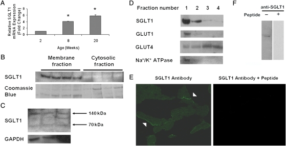
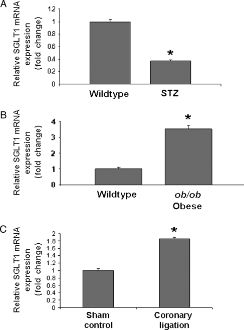
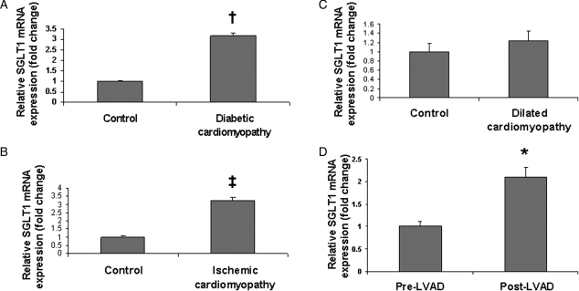
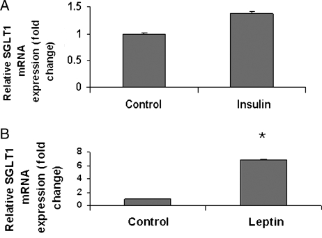
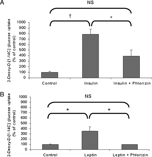
Methods and results: SGLT1 was largely localized to the cardiac myocyte sarcolemma. Changes in SGLT1 expression were observed in disease states in both humans and mouse models. SGLT1 expression was upregulated two- to three-fold in type 2 diabetes mellitus and myocardial ischaemia (P < 0.05). In humans with severe heart failure, functional improvement following implantation of left ventricular assist devices led to a two-fold increase in SGLT1 mRNA (P < 0.05). Acute administration of leptin to wildtype mice increased cardiac SGLT1 expression approximately seven-fold (P < 0.05). Insulin- and leptin-stimulated cardiac glucose uptake was significantly (P < 0.05) inhibited by phlorizin, a specific SGLT1 inhibitor.

Conclusion: Our data suggest that cardiac SGLT1 expression and/or function are regulated by insulin and leptin, and are perturbed in disease. This is the first study to examine the regulation of cardiac SGLT1 expression by insulin and leptin and to determine changes in SGLT1 expression in cardiac disease.


Figures

Figure 1
Figure 1
SGLT1 is expressed in murine and human cardiac myocytes. (A) Relative SGLT1 mRNA expression was assessed by QPCR in hearts harvested from male WT FVB mice at ages 2, 8, and 20 weeks (n = 5 per group). Data are expressed as mean ± SE. *P < 0.01 relative to 2-week-old hearts. (B) A representative immunoblot of membrane and cytosolic fractions of murine cardiac protein showed that SGLT1 was present only in the membrane fraction. Coomassie blue staining of the protein gel was used to document the relative quantity of protein loaded for the immunoblot. (C) A representative immunoblot of total human cardiac protein showed the presence of two (70 and 140 kDa) SGLT1 bands in all lanes, and an intermediate band in the rightmost lane. An immunoblot of GAPDH was used to document the relative quantity of protein loaded. (D) A representative immunoblot of murine cardiac protein fractions derived on a sucrose gradient showed colocalization of SGLT1, GLUT1, and Na+/K+ ATPase (a marker for the sarcolemma). (E) Immunofluorescence microscopy showed that SGLT1 was predominantly localized to the sarcolemma of cardiac myocytes from 8-week-old male WT FVB mice (left, arrowheads), a staining pattern that was significantly decreased by pre-incubation of the antibody with the immunizing peptide. (F) Further to demonstrate anti-murine SGLT1 antibody specificity, the SGLT1 band visualized by immunoblot was completely competed off by pre-incubation of the antibody with the peptide.
Figure 2
Figure 2
Perturbations in levels of cardiac SGLT1 mRNA expression as measured by QPCR in diseased murine hearts relative to control. Cardiac SGLT1 expression was significantly (A) decreased in streptozotocin (STZ) treated (type 1 diabetic) FVB mice, (B) increased in ob/ob (type 2 diabetic) mice, and (C) increased in WT C57BL/6J mice after coronary artery ligation. n = 4–6 per group. Data are expressed as mean ± SE. *P < 0.05.
Figure 3
Figure 3
Perturbations in levels of cardiac SGLT1 mRNA expression as measured by QPCR in diseased human hearts. Significant increases in SGLT1 expression were observed in subjects with end-stage cardiomyopathy secondary to (A) long-standing type 2 diabetes and (B) coronary artery disease, relative to age- and sex-matched control subjects. (C) No change in SGLT1 expression was observed in idiopathic dilated cardiomyopathy. (D) In failing hearts, implantation of left ventricular assist devices (LVAD) resulted in an increase in SGLT1 expression, which correlated with improved contractile function. Paired samples from the same subjects were used. n = 5–6 per group. Data are expressed as mean ± SE. *P < 0.05; P < 0.01; P < 0.001 relative to control or baseline.
Figure 4
Figure 4
Acute effects of (A) insulin in WT FVB mice and (B) leptin in WT C57BL/6J mice on cardiac SGLT1 mRNA expression as measured by QPCR 30 min following administration. Leptin, but not insulin, significantly increased SGLT1 expression. n = 3–5 per group. Data are expressed as mean ± SE. *P < 0.05; NS, not significant.
Figure 5
Figure 5
Increased cardiac glucose uptake in WT FVB mice in response to exogenous insulin and leptin administration is dependent on SGLT1. (A) Increased glucose uptake observed after insulin administration was inhibited by phlorizin, a SGLT1 inhibitor. (B) Increased glucose uptake observed after leptin administration was completely inhibited by phlorizin (n = 3 per group). Data are expressed as mean ± SE. *P < 0.05; P < 0.01; NS, not significant.

References

    1. Stanley WC, Recchia FA, Lopaschuk GD. Myocardial substrate metabolism in the normal and failing heart. Physiol Rev. 2005;85:1093–1129. - PubMed
    1. Stanley WC, Lopaschuk GD, Hall JL, McCormack JG. Regulation of myocardial carbohydrate metabolism under normal and ischaemic conditions. Potential for pharmacological interventions. Cardiovasc Res. 1997;33:243–257. - PubMed
    1. Schwenk RW, Luiken JJ, Bonen A, Glatz JF. Regulation of sarcolemmal glucose and fatty acid transporters in cardiac disease. Cardiovasc Res. 2008;79:249–258. - PubMed
    1. Wright EM, Hirsch JR, Loo DD, Zampighi GA. Regulation of Na+/glucose cotransporters. J Exp Biol. 1997;200:287–293. - PubMed
    1. Turk E, Zabel B, Mundlos S, Dyer J, Wright EM. Glucose/galactose malabsorption caused by a defect in the Na+/glucose cotransporter. Nature. 1991;350:354–356. - PubMed

Publication types

MeSH terms