. 2009 Jul 1;122(Pt 13):2311-21.
doi: 10.1242/jcs.046557. Epub 2009 Jun 9.

Distinct roles for isoforms of the catalytic subunit of class-IA PI3K in the regulation of behaviour of murine embryonic stem cells

Affiliations

Distinct roles for isoforms of the catalytic subunit of class-IA PI3K in the regulation of behaviour of murine embryonic stem cells

Emmajayne Kingham et al. J Cell Sci. .

Abstract

Self-renewal of embryonic stem cells (ESCs) is essential for maintenance of pluripotency, which is defined as the ability to differentiate into any specialised cell type comprising the adult organism. Understanding the mechanisms that regulate ESC self-renewal and proliferation is required before ESCs can fulfil their potential in regenerative therapies, and murine ESCs (mESCs) have been widely used as a model. Members of the class-IA phosphoinositide 3-kinase (PI3K) family of lipid kinases regulate a variety of physiological responses, including cell migration, proliferation and survival. PI3Ks have been reported to regulate both proliferation and self-renewal of mESCs. Here we investigate the contribution of specific class-IA PI3K isoforms to the regulation of mESC fate using small-molecule inhibitors with selectivity for particular class-IA PI3K catalytic isoforms, and siRNA-mediated knockdown. Pharmacological inhibition or knockdown of p110beta promoted mESC differentiation, accompanied by a decrease in expression of Nanog. By comparison, pharmacological inhibition or siRNA-mediated knockdown of p110alpha had no effect on mESC self-renewal per se, but instead appeared to reduce proliferation, which was accompanied by inhibition of leukaemia inhibitory factor (LIF) and insulin-induced PI3K signalling. Our results suggest that PI3Ks contribute to the regulation of both mESC pluripotency and proliferation by differential coupling to selected p110 catalytic isoforms.


Figures
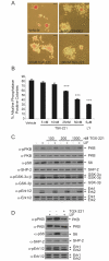
Fig. 1.
Fig. 1.
Pharmacological inhibition of p110β leads to a decrease in self-renewal of murine ESCs. (A) Murine ESCs (mESCs), cultured in the presence of LIF and serum, were treated for 4 days with LY294002 or TGX-221 at the doses shown or with vehicle alone (DMSO). Self-renewal was assessed by staining for alkaline-phosphatase activity and representative colonies are shown. Scale bars: 100 μm. (B) The average percentage of alkaline-phosphatase-positive, self-renewing colonies in each condition are shown with s.e.m. (n=4). ANOVA and Dunnett's post-hoc test were applied; ***P<0.001. (C) Cells were treated with TGX-221 for 30 minutes prior to stimulation with 103 U/ml LIF for 10 minutes. Cell lysates were resolved by SDS-PAGE and immunoblotting carried out using the antibodies indicated to detect PI3K downstream signalling. (D) Cells were cultured for 4 days in the presence or absence of LIF or in the presence of LIF and 100 nM TGX-221 and immunoblotting carried out as above.
Fig. 2.
Fig. 2.
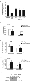
Knockdown of Pik3cb reduces self-renewal and decreases Nanog expression. mESCs were transfected with Pik3cb (p110β), Nanog or non-targeting (NT) Smartpool siRNAs and harvested 72 hours later. (A) Quantitative RT-PCR was conducted on quadruplicate samples to measure knockdown of Pik3cb RNA and levels of RNA encoding p110β were normalised relative to levels of β-actin RNA. Representative data from four independent experiments are shown with s.d.; *P<0.05 following a Student's t-test. (B) Quantitative RT-PCR was conducted to detect knockdown of Nanog RNA. Representative data are shown with s.d.; *P<0.05 and **P<0.01 following a Student's t-test. (C) Following transfection, cells were plated for 4 days and self-renewal assessed by alkaline-phosphatase staining. The average percentage of alkaline-phosphatase-positive, self-renewing colonies in each condition are shown with s.e.m. (n=4); *P<0.05 following a Student's t-test. The number of total colonies formed in each condition did not vary significantly. (D) Assessment of self-renewal by alkaline-phosphatase staining is depicted by the percentage of pure alkaline-phosphatase colonies (compact round red-stained colonies) with s.e.m. (n=4); * P<0.05 following a Student's t-test. (E) Immunoblotting was used to detect (i) Nanog and Oct4 protein levels or (ii) p85 protein levels, with SHP-2 reprobing used to confirm equal loading.
Fig. 3.
Fig. 3.
siRNA-mediated knockdown of p110β expression reduces self-renewal. mESCs were transfected with Pik3cb-targeting siRNAs or with Negative Control Silencer Select siRNA (Ambion). (A) Following transfection, cells were plated for 4 days and self-renewal assessed by alkaline-phosphatase staining. The average percentage of alkaline-phosphatase-positive colonies are shown, plus s.e.m. (n=4); *P<0.05 and **P<0.01 following a Student's t-test. The number of total colonies formed in each condition did not vary significantly. (B,C) Following transfection, cells were re-plated for a further 4 days then harvested for RNA analysis. Quantitative RT-PCR was conducted to detect knockdown of Pik3cb RNA (B) or expression of Nanog (C). Representative data, with s.d., are shown; *P<0.05 and **P<0.01 following a Student's t-test. The percentage of knockdowns, compared with the negative control, are indicated. (D) Immunoblots detecting expression of Nanog and p85 proteins in mESCs after transfection and 4 days of culture are shown and the position of proteins indicated. Blots were stripped and reprobed with antibodies detecting SHP-2 to assess loading.
Fig. 4.
Fig. 4.
A minor role for p110δ in mESC self-renewal. (A) mESCs were treated with a p110δ inhibitor, IC87114, for 4 days and self-renewal assessed by staining for alkaline-phosphatase activity. The average percentage of alkaline-phosphatase-positive, self-renewing colonies in each condition are shown with s.e.m. (n=4); *P<0.05 following a Student's t-test. The number of total colonies formed in each condition did not vary significantly. (B) Quantitative RT-PCR was conducted to detect knockdown of Pik3cd RNA following transfection with Pik3cd-targeting siRNA or with non-targeting siRNA. Representative data is shown with s.d. and the percentage of knockdowns, compared with control, are indicated. (C) Following transfection, cells were plated for 4 days and self-renewal assessed by alkaline-phosphatase staining. The average percentage of alkaline-phosphatase-positive, self-renewing colonies in each condition are shown with s.e.m. The number of total colonies formed in each condition did not vary significantly. (D) Expression of Nanog was assessed by quantitative RT-PCR following knockdown of Pik3cd. Representative data is shown with s.d. (E) Immunoblotting was carried out to detect p85 protein levels, with SHP-2 reprobing used to confirm equal loading.
Fig. 5.
Fig. 5.
p110α does not regulate mESC self-renewal. mESCs were transfected with Pik3ca-targeting (p110α) or with non-targeting (NT) siRNAs. (A) Quantitative RT-PCR of quadruplicate samples was conducted to detect knockdown of Pik3ca RNA. The averages, with s.d., are shown and are representative of four independent experiments. *P<0.05 following a Student's t-test. (B) Following transfection, cells were plated for 4 days and self-renewal assessed by alkaline-phosphatase staining. The average percentage of alkaline-phosphatase-positive, self-renewing colonies in each condition are shown with s.e.m. (n=4). The total number of colonies formed in each condition did not vary significantly. (Ci) Semi-quantitative RT-PCR was used to assess knockdown of RNA encoding p110α, Nanog or Rex1 following transfection with the indicated siRNAs. (Cii) Immunoblotting was used to detect Nanog, p85 and SHP-2 protein levels. (D) Nanog expression was also assessed by quantitative RT-PCR as in B. (E,F) mESCs were treated for 4 days with the indicated concentrations of LY294002, compound 15e (E), PIK-75 (F) or with vehicle alone (DMSO). The average number of self-renewing alkaline-phosphatase-positive and non-self-renewing colonies in each condition is shown with s.e.m. (15e, n=5; PIK-75, n=3); *P<0.05 following a Student's t-test. (G) Following treatment with 15e or PIK-75, colonies were fixed and stained for alkaline phosphatase. Representative colonies are depicted. Scale bars: 100 μm.
Fig. 6.
Fig. 6.
p110α appears to be coupled to proliferation, insulin signalling and LIF signalling in mESCs. (A) To assess cell proliferation, viable cell numbers were determined in triplicate at 24-hour intervals for up to 4 days. The average number of cells per dish, ± s.d., are shown for each treatment. (B,C) Following 30 minutes pre-treatment with either (i) 15e or (ii) PIK-75 at the doses indicated, mESCs were stimulated with 10 μg/ml insulin for 5 minutes (B) or 103 U/ml LIF for 10 minutes (C). Signalling downstream of PI3K was assessed by SDS-PAGE and immunoblotting with the antibodies indicated. Data, representative of three independent experiments, are shown.
Fig. 7.
Fig. 7.
p110α inhibition does not prevent loss of self-renewal or block mESC differentiation. (A) mESCs were treated with the p110α inhibitors 15e or PIK-75 for 4 days, at the doses indicated, in the presence or absence of LIF. The average percentage of alkaline-phosphatase-positive colonies, with s.e.m. (n=3), is shown, ***P<0.001 following a Student's t-test. (B) The average number of self-renewing alkaline-phosphatase-positive and non-self-renewing colonies in each condition is shown with s.e.m. (n=3). (C) mESCs cultured in the presence of LIF were treated with TGX-221 for 4 days in the presence or absence of 600 nM 15e. Self-renewal was assessed by staining for alkaline-phosphatase activity and the average percentage of alkaline-phosphatase-positive colonies, with s.e.m., are shown (n=4). ANOVA (P=0.002) and Dunnett's post-hoc test were applied; **P<0.01.

Similar articles

Cited by

References

    1. Aksoy, I., Sakabedoyan, C., Bourillot, P. Y., Malashicheva, A. B., Mancip, J., Knoblauch, K., Afanassieff, M. and Savatier, P. (2007). Self-renewal of murine embryonic stem cells is supported by the serine/threonine kinases pim-1 and pim-3. Stem Cells 25, 2996-3004. - PubMed
    1. Armstrong, L., Hughes, O., Yung, S., Hyslop, L., Stewart, R., Wappler, I., Peters, H., Walter, T., Stojkovic, P., Evans, J. et al. (2006). The role of PI3K/AKT, MAPK/ERK and NFkappabeta signalling in the maintenance of human embryonic stem cell pluripotency and viability highlighted by transcriptional profiling and functional analysis. Hum. Mol. Gen. 15, 1894-1913. - PubMed
    1. Bi, L., Okabe, I., Bernard, D. J., Wynshaw-Boris, A. and Nussbaum, R. L. (1999). Proliferative defect and embryonic lethality in mice homozygous for a deletion in the p110alpha subunit of phosphoinositide 3-kinase. J. Biol. Chem. 274, 10963-10968. - PubMed
    1. Bi, L., Okabe, I., Bernard, D. J. and Nussbaum, R. L. (2002). Early embryonic lethality in mice deficient in the p110beta catalytic subunit of PI 3-kinase. Mamm. Genome 13, 169-172. - PubMed
    1. Boeuf, H., Hauss, C., DeGraeve, F., Baran, N. and Kedinger, C. (1997). Leukemia inhibitory factor-dependent transcriptional activation in embryonic stem cells. J. Cell Biol. 138, 1207-1217. - PMC - PubMed

Publication types

MeSH terms