

Case Reports
doi: 10.3174/ajnr.A1682.
Epub 2009 Jun 9.
Tuberculosis involving bilateral middle cerebellar peduncles
- PMID: 19509071
- PMCID: PMC7051619
- DOI: 10.3174/ajnr.A1682
Item in Clipboard
Case Reports
Tuberculosis involving bilateral middle cerebellar peduncles
AJNR Am J Neuroradiol.
2009 Sep.
No abstract available
Figures
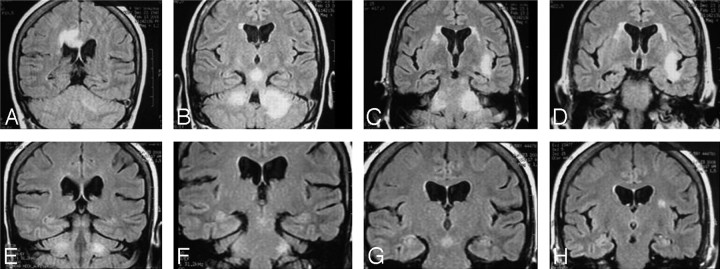
Coronal FLAIR MR images show hyperintense lesions in the cingulate cortex area involving the corpus callosum (A), central midbrain (B), asymmetric bilateral MCP (B and C), and left perisylvian area (C and D). Five months later, images show minimal hyperintense lesions in both the MCP (E and F), central midbrain (G), and left perisylvian area (H).
References
-
- Pui MH, Memon WA. Magnetic resonance imaging findings in tuberculous meningoencephalitis. Can Assoc Radiol J 2001; 52: 43– 49 - PubMed
-
- Bernaerts A, Vanhoenacker F, Parizel PM, et al. Tuberculosis of the central nervous system: overview of neuroradiological findings. Eur Radiol 2003; 13: 1876– 90 - PubMed
-
- Kais N, Allani R, Abdelmalek R, et al. Value of magnetic resonance imaging in central nervous system tuberculosis. Presse Med 2008; 37( 4 pt 2): 634– 42 - PubMed
Publication types
MeSH terms
LinkOut - more resources
Full Text Sources
Medical