. 2009 Jul 1;69(13):5522-30.
doi: 10.1158/0008-5472.CAN-09-0266. Epub 2009 Jun 9.

Malignant B cells skew the balance of regulatory T cells and TH17 cells in B-cell non-Hodgkin's lymphoma

Affiliations

Malignant B cells skew the balance of regulatory T cells and TH17 cells in B-cell non-Hodgkin's lymphoma

Zhi-Zhang Yang et al. Cancer Res. .

Abstract

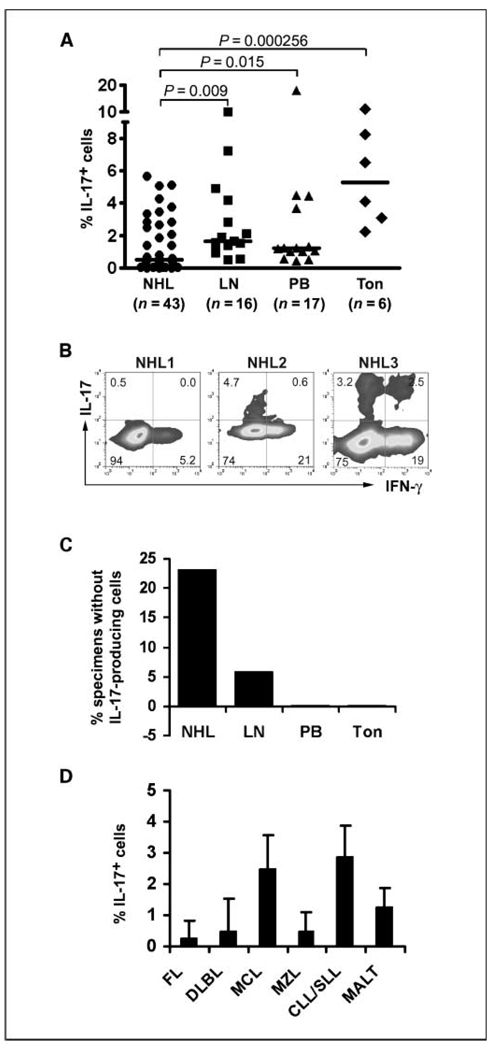
Using biopsy specimens from patients with B-cell non-Hodgkin's lymphoma, we observed a significantly low frequency of T(H)17 cells, including several samples with no detectable amount of interleukin (IL)-17-producing cells present in the tumor microenvironment. We found that, in the absence of lymphoma B cells, treatment with IL-1beta/IL-6 or lipopolysaccharide (LPS) enhanced IL-17 expression in CD4(+) T cells and this enhancement was attenuated when CD4(+) T cells were cocultured with lymphoma B cells. Blockade of CD27-CD70 or CD28-CD80/86 interactions by anti-CD70 or anti-CD80/86 antibodies restored LPS-mediated induction of IL-17 expression in CD4(+) T cells cocultured with lymphoma B cells. Because a subset of lymphoma B cells express IL-2 and given that IL-2 signaling is critically important in the development of regulatory T (T(reg)) cells, we tested the role of IL-2 signaling in T(H)17 cell development. We found that treatment with anti-IL-2 antibody to interrupt IL-2 signaling significantly inhibited Foxp3 expression in CD4(+) T cells. In contrast, interruption of IL-2 signaling up-regulated IL-17 expression in CD4(+) T cells and restored lymphoma-mediated down-regulation of IL-17-producing cells. Furthermore, the reversal of T(reg) cell activity by LPS or CpG-A resulted in an enhancement of IL-17-producing cells. Taken together, our study indicated that lymphoma B cells play an important role in skewing the balance between T(reg) and T(H)17 cells resulting in the establishment of a profoundly inhibitory tumor microenvironment.


Conflict of interest statement

Disclosure of Potential Conflicts of Interest

No potential conflicts of interest were disclosed.

Figures

Figure 1
Figure 1
Frequency of TH17 cells in B-cell non-Hodgkin’s lymphoma biopsy specimens. A, frequency of CD4+ IL-17–producing cells from B-cell non-Hodgkin’s lymphoma (NHL), benign lymph node (LN), peripheral blood from healthy donors (PB), and benign tonsil tissues (Ton). Cells were cultured in anti-CD3 antibody-coated plates with anti-CD28 antibody for 3 days and restimulated with phorbol 12-myristate 13-acetate/ionomycin in the presence of protein transport inhibitor brefeldin A for 5 h. Cells were subjected to intracellular staining with fluorochrome-conjugated antibodies specific to CD3, CD4, IL-17, and IFN-γ. The frequency of TH17 cells was measured as % CD4+ IL-17–producing T cells. B, IL-17 and IFN-γ expression in CD4+ T cells from three representative specimens. C, numbers of specimens without detectable IL-17–producing cells. D, frequency of CD4+ IL-17–producing cells from indicated histologic types of B-cell non-Hodgkin’s lymphoma. FL, follicular lymphoma; DLBCL, diffuse large B-cell lymphoma; MCL, mantle cell lymphoma; MZL, marginal zone lymphoma; CLL/SLL, small lymphocytotic lymphoma; MALT, mucosa-associated lymphoid tissue.
Figure 2
Figure 2
Phenotype of TH17 cells. A, left, IL-17 expression in CD4+ or CD8+ T cells; right, summarized data on the frequency of IL-17–producing cells from different types of cells. B, IL-17 expression in CD4+CD45RA+ or CD4+CD45RO+ T cells determined by flow cytometry (left) and ELISA (right). C, IL-17 expression in CD4+CCR6+ or CD4+CCR6- T cells determined by flow cytometry (left) and ELISA (right).
Figure 3
Figure 3
Cytokine regulation of TH17 cells. A, IL-17 expression in CD4+ T cells treated with indicated cytokine. B, left, summary of IL-17 induction in CD4+ T cells incubated without (NIL) or with IL-1β/IL-6 (CKs). Horizontal bars, median. Right, summary of IL-17 concentration in culture supernatants in CD4+ T cells incubated without or with IL-1β/IL-6. C, IL-17 expression in CD4+ T cells treated without or with IL-1β/IL-6 in the absence or presence of TGF-β. D, IL-17 expression in CD4+ T cells treated without or with IL-1β/IL-6 in the absence or presence of anti-IFN-γ antibody.
Figure 4
Figure 4
Effect of malignant B cells on TH17 cells. A, IL-17 expression in CD4+ T cells from non-Hodgkin’s lymphoma tissue without depletion (U, unsorted) or with depletion (D, CD19-depleted) of B cells. Right, summarized data (n = 8 patient samples). B, IL-17 expression in CD4+ T cells isolated from non-Hodgkin’s lymphoma tissue and cocultured without (top) or with (bottom) CD19+ lymphoma cells (LB) in the absence (NIL) or presence of IL-1β/IL-6 (CKs) or LPS. C, IL-17 and IFN-γ expression in CD4+ T cells cocultured without or with CD19+ lymphoma cells in the absence or presence of LPS plus blocking antibody specific to CD70 or CD80 or CD86.
Figure 5
Figure 5
Effect of IL-2 signaling on TH17 cells. A, Foxp3 expression in CD4+ T cells isolated from tumor biopsy specimens treated without or with TGF-β in the absence or presence of anti-IL-2 antibody (n = 3). B, IL-17 and IFN-γ expression in CD4+ T cells treated without or with IL-1β/IL-6 in the presence or absence of anti-IL-2 (top), anti-IL-2Rα (middle), or anti-IL-2Rβ (bottom) antibody (n = 5). Each panel represents a patient specimen. C, IL-2 expression in CD19+ cells activated without (Unstim) or with LPS and phorbol 12-myristate 13-acetate/ionomycin (Stim; n = 4non-Hodgkin’s lymphoma samples). D, IL-17 and IFN-γ expression in CD4+ T cells cocultured with CD19+ lymphoma B cells in the absence or presence of IL-1β/IL-6 plus blocking antibody specific to IL-2 (aIL-2) or isotype control (IgG; n = 3 non-Hodgkin’s lymphoma samples).
Figure 6
Figure 6
Reciprocal regulation of TH17 and Treg cells in B-cell non-Hodgkin’s lymphoma. A, IL-17 and IFN-γ expression in CD4+ T cells treated without or with IL-1β/IL-6 or LPS or CpG-A or non-CpG-A (n = 3 non-Hodgkin’s lymphoma samples). B, Foxp3 (top) or IL-17 (bottom) expression in CD4+ T cells from a representative specimen treated without or with TGF-β or TGF-β plus IL-23 (n = 4non-Hodgkin’s lymphoma samples). C, a paired representative sample showing IL-17 and Foxp3 expression in tonsil CD4+ T cells from a normal individual or a patient with B-cell non-Hodgkin’s lymphoma. D, summary of Foxp3 (open dots) and IL-17 (dark dots) expression in CD4+ T cells in response to different stimulation. The induction of Foxp3 or IL-17 expression in CD4+ T cells was converted to logarithm number. IL-2 signaling interruption: CD4+ T cells were treated with anti-IL-2, IL-2Rα, or IL-2Rβ antibody; TGF-β: CD4+ T cells were treated with TGF-β; non-Hodgkin’s lymphoma B cells: CD4+ T cells were cocultured with non-Hodgkin’s lymphoma cells in the presence of IL-1β/IL-6.

References

    1. Harrington LE, Hatton RD, Mangan PR, et al. Interleukin 17-producing CD4+ effector T-cells develop via a lineage distinct from the T helper type 1and 2 lineages. Nat Immunol. 2005;6:1123–1132. - PubMed
    1. Park H, Li Z, Yang XO, et al. A distinct lineage of CD4 T-cells regulates tissue inflammation by producing interleukin 17. Nat Immunol. 2005;6:1133–1141. - PMC - PubMed
    1. Bettelli E, Carrier Y, Gao W, et al. Reciprocal developmental pathways for the generation of pathogenic effector TH17 and regulatory T-cells. Nature. 2006;441:235–238. - PubMed
    1. Sakaguchi S. Naturally arising CD4+ regulatory T-cells for immunologic self-tolerance and negative control of immune responses. Ann Rev Immunol. 2004;22:531–562. - PubMed
    1. Chen W, Jin W, Hardegen N, et al. Conversion of peripheral CD4+CD25− naive T-cells to CD4+CD25+ regulatory T-cells by TGF-β induction of transcription factor Foxp3. J Exp Med. 2003;198:1875–1886. - PMC - PubMed

Publication types

MeSH terms