

Orally active alpha-tocopheryloxyacetic acid suppresses tumor growth and multiplicity of spontaneous murine breast cancer
- PMID: 19509249
- PMCID: PMC3693733
- DOI: 10.1158/1535-7163.MCT-08-1079
Orally active alpha-tocopheryloxyacetic acid suppresses tumor growth and multiplicity of spontaneous murine breast cancer
Abstract
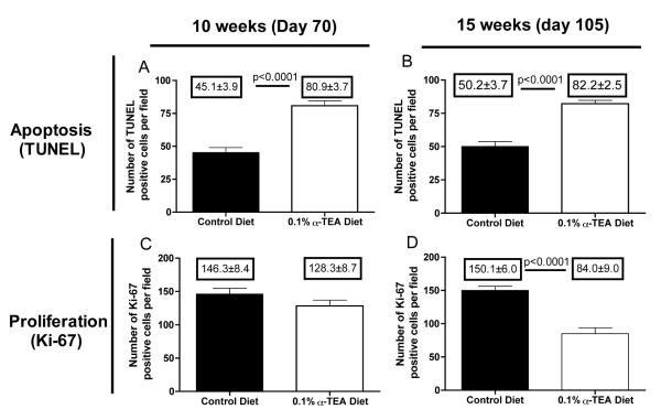
We recently demonstrated the antitumor efficacy of orally administered alpha-tocopheryloxyacetic acid (alpha-TEA), a redox silent and nonhydrolyzable derivative of naturally occurring vitamin E. In order to move alpha-TEA closer to the clinic to benefit patients with breast cancer, the present study had two goals. First, to determine the minimal effective treatment dose; and second, to test the efficacy of dietary administration of alpha-TEA in the clinically relevant MMTV-PyMT mouse model of spontaneous breast cancer that more closely resembles human disease. The minimal effective dose of alpha-TEA was evaluated in the transplantable 4T1 tumor model and we show a dose-dependent decrease of primary tumor growth and reduction of metastatic spread to the lung. Six-week-old MMTV-PyMT mice were treated with oral alpha-TEA for 9 weeks, with no apparent signs of drug toxicity. The alpha-TEA treatment delayed tumor development and significantly slowed tumor progression, resulting in a 6-fold reduction of the average cumulative tumor size. In addition, oral alpha-TEA caused an 80% reduction in spontaneous metastases. In situ analysis of tumor tissue identified apoptosis as an important mechanism of alpha-TEA-mediated tumor suppression in addition to inhibition of tumor cell proliferation. This study shows, for the first time, the ability of orally administered alpha-TEA to delay tumor onset and to inhibit the progression and metastatic spread of a clinically relevant model of spontaneous breast cancer. Our finding of the high efficacy in this tumor model highlights the translational potential of oral alpha-TEA therapy.
Figures
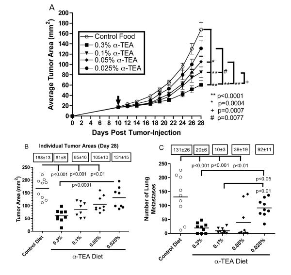
 ,~17 mm2, day 10 post-tumor cell injection), mice received α-TEA in the diet for 18 days. Untreated mice received control diet throughout the study. (A) The values represent the mean tumor areas ± SEM of 9 mice per group. To compare tumor growth rates, growth curves were transformed to linearity and linear regression analysis was used to determine slopes that were then compared by t-test. (B) The values represent tumor areas of individual mice on day 28 post-tumor injection. Boxed numbers are mean tumor areas ± SEM. Differences of the mean tumor areas were determined by ANOVA including Tukey-Kramer post tests for multiple comparisons. (C) Effect of dietary delivery of
α-TEA on tumor spread. BALB/c mice with implanted 4T1 mammary tumors from the above study were sacrificed on day 28 post-tumor cell injection. To determine the number of pulmonary metastases, lungs were inflated with India ink and removed, and the surface lung metastases were counted. Boxed numbers are mean number of lung metastases ± SEM. Differences of the mean number of lung metastases were determined by ANOVA including Tukey-Kramer post tests for multiple comparisons.
,~17 mm2, day 10 post-tumor cell injection), mice received α-TEA in the diet for 18 days. Untreated mice received control diet throughout the study. (A) The values represent the mean tumor areas ± SEM of 9 mice per group. To compare tumor growth rates, growth curves were transformed to linearity and linear regression analysis was used to determine slopes that were then compared by t-test. (B) The values represent tumor areas of individual mice on day 28 post-tumor injection. Boxed numbers are mean tumor areas ± SEM. Differences of the mean tumor areas were determined by ANOVA including Tukey-Kramer post tests for multiple comparisons. (C) Effect of dietary delivery of
α-TEA on tumor spread. BALB/c mice with implanted 4T1 mammary tumors from the above study were sacrificed on day 28 post-tumor cell injection. To determine the number of pulmonary metastases, lungs were inflated with India ink and removed, and the surface lung metastases were counted. Boxed numbers are mean number of lung metastases ± SEM. Differences of the mean number of lung metastases were determined by ANOVA including Tukey-Kramer post tests for multiple comparisons.
References
-
- Lawson KA, Anderson K, Menchaca M, et al. Novel vitamin E analogue decreases syngeneic mouse mammary tumor burden and reduces lung metastasis. Moleculaar Cancer Therapy. 2003;2:437–44. - PubMed
-
- Hahn T, Szabo L, Gold M, Ramanathapuram L, Hurley LH, Akporiaye ET. Dietary Administration of the Proapoptotic Vitamin E Analogue {alpha}-Tocopheryloxyacetic Acid Inhibits Metastatic Murine Breast Cancer. Cancer Research. 2006;66:9374–8. - PubMed
-
- Neuzil J, Tomasetti M, Mellick AS, et al. Vitamin E analogues: a new class of inducers of apoptosis with selective anti-cancer effects. Current Cancer Drug Targets. 2004;4:355–72. - PubMed
Publication types
MeSH terms
Substances
Grants and funding
LinkOut - more resources
Full Text Sources
Other Literature Sources
