. 2009 Jun;8(6):1646-54.
doi: 10.1158/1535-7163.MCT-08-1084. Epub 2009 Jun 9.

Length of mitotic arrest induced by microtubule-stabilizing drugs determines cell death after mitotic exit

Affiliations

Length of mitotic arrest induced by microtubule-stabilizing drugs determines cell death after mitotic exit

Michael E Bekier et al. Mol Cancer Ther. 2009 Jun.

Abstract

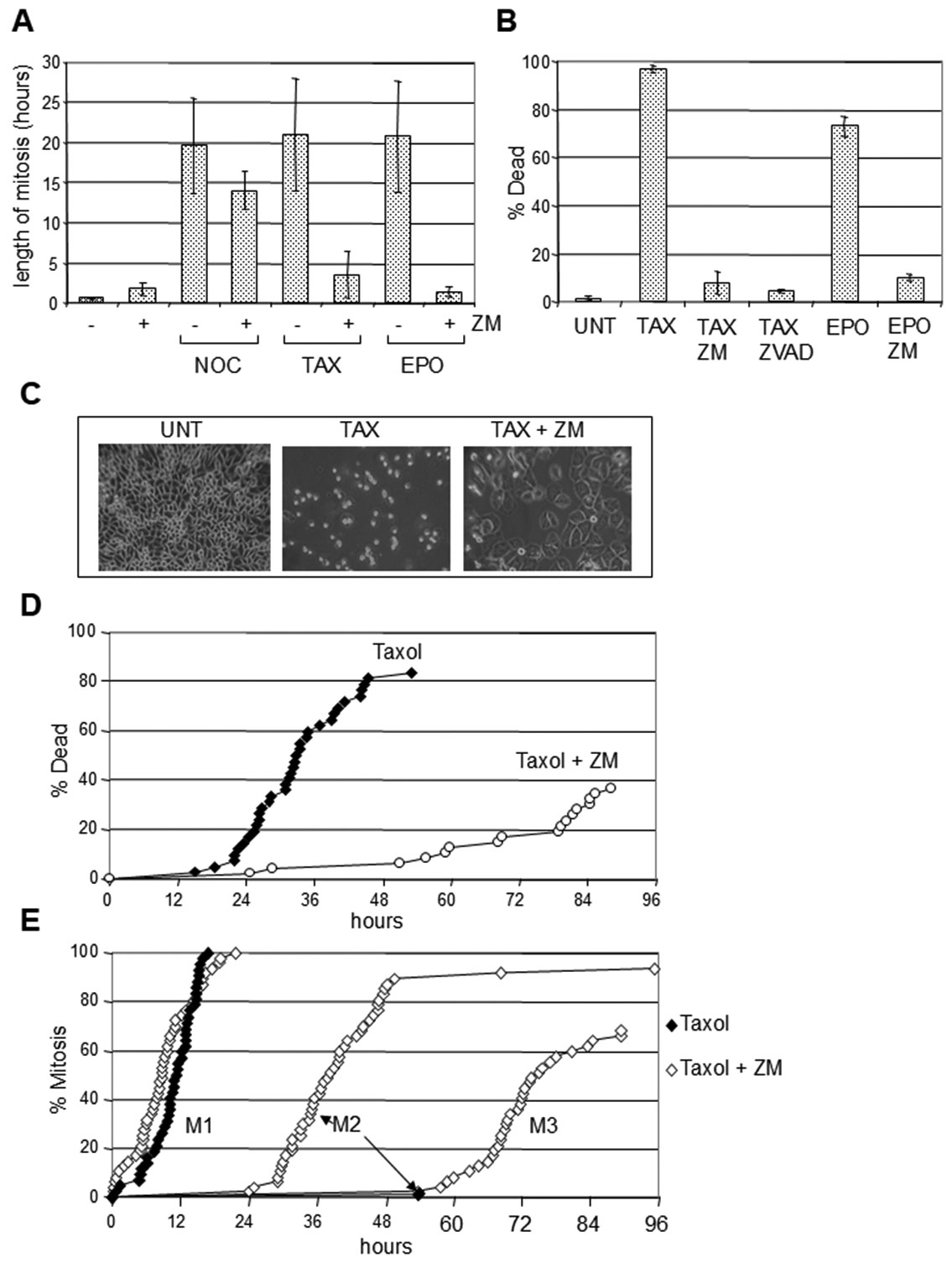
Cell death induced by agents that disrupt microtubules can kill cells by inducing a prolonged mitotic block. This mitotic block is dependent on the spindle assembly checkpoint, a surveillance system that ensures the bipolar attachment of chromosomes to the mitotic spindle before the onset of anaphase. Under some conditions, the spindle assembly checkpoint can become weakened, allowing cells to exit mitosis despite the presence of chromosomes that are not properly attached to the mitotic spindle. Here, we use an Aurora kinase inhibitor to drive mitotic exit and test the effect of mitotic arrest length on death in the subsequent interphase. Cells that are blocked in mitosis for >15 h die shortly after exiting from mitosis, whereas cells that exit after being blocked for <15 h show variable fates, with some living for days after exiting mitosis. Cells blocked in mitosis by either Taxol or epothilone B are acutely sensitive to the death ligand tumor necrosis factor-related apoptosis-inducing ligand, suggesting that prolonged mitosis allows the gradual accumulation of internal death signals, rendering cells hypersensitive to additional prodeath cues. Death under these conditions is initiated while cyclin B1 is still present, indicating that cells are in mitosis. Our experiments suggest that there is a point of no return during prolonged mitotic block after which mitotic exit can no longer block death.


Figures

Figure 1.
Figure 1.
ZM447439 abrogates mitotic arrest and inhibits cell death by microtubule disruptive drugs. HelaM cells were exposed to 10μM Taxol (TAX), 20nM Epothilone B (EPO), or to 0.2μg/ml Nocodazole in the presence or absence of 2.5μM ZM447439 (ZM) or 40μM Z-VAD-FMK (ZVAD). (A) Length of mitosis as determined by time-lapse microscopy. In the case of cells treated with Taxol, Nocodazole or Epothilone B alone, mitosis ended when the cells died. In other treatments, mitosis ended when the cells entered interphase. (B) Cell death as assessed by trypan blue exclusion. HelaM cells were exposed to the indicated drugs for 48 hr before staining with trypan blue. (C) Examples of cells exposed to Taxol for 48 hr in the presence or absence of 2.5μM ZM447439. (D) Cell death as measured by time-lapse microscopy. HelaM cells exposed to Taxol (10μM) in the presence or absence of ZM447439 (ZM; 2.5μM) were tracked and cumulative events are shown. (E) Mitosis as assessed by time-lapse microscopy. HelaM cells treated as in “D” attempted mitosis several times (M1, M2 and M3). Under these conditions, cells were unable to divide and those that attempted mitosis several times became giant and multinucleated.
Figure 2.
Figure 2.
Mitotic exit followed by cell death after treatment with Nocodazole and ZM447439. HelaM cells simultaneously treated with 0.2μg/ml Nocodazole and 2.5μM ZM447439 were analyzed by time-lapse microscopy. (A) Examples of cells entering mitosis, exiting mitosis and then dieing. (B) Cumulative mitotic entry, mitotic exit and death.
Figure 3.
Figure 3.
Length of mitotic arrest determines cell death upon mitotic exit. HelaM cells were exposed to 10μM Taxol for 24 hr, followed by 2.5μM ZM447439. Entry into mitosis during the Taxol treatment was tracked by time- lapse microscopy. Tracking was continued on the same field of cells after the addition of ZM447439 to determine mitotic exit and cell death. (A) Mitotic exit induced by ZM447439. The length of time for cells to exit mitosis after exposure to ZM447439 is compared to how long they had been previously blocked in mitosis in the presence of Taxol. (B) Fate of cells after mitotic exit as a function of the length of mitotic block. The time of cell death is compared to the length of the previous mitotic arrest. Some cells did not die during the entire length of the experiment (white triangles) and several exited mitosis before ZM447439 was added (white circles). All cells in the field did exit mitosis after treatment with ZM447439.
Figure 4.
Figure 4.
Inhibition of Taxol/Trail killing by ZM447439. HelaM cells were exposed to the drugs indicated and cell viability determined 48 hr later using the MTS assay as described in Materials and Methods. TAX: Taxol; ZM: ZM4474379; Trail was added at 10ng/ml.
Figure 5.
Figure 5.
Cell death in response to Trail in combination with Taxol or Epothilone B. HelaM cells were exposed to the indicated drugs and cell fates determined by time-lapse microscopy. (A) Simultaneous exposure to Taxol and Trail. HelaM cells were exposed to 0.5 μM Taxol and 10ng/ml Trail and then immediately tracked by time-lapse microscopy to correlate mitosis with cell death. 89% of cells died during filming, with the majority dieing during mitosis (79% of total). Time of death is compared to time of entry into mitosis. (B) Addition of Trail after Taxol. HelaM cells were exposed to 10 μM Taxol for 18 hr after which 10ng/ml Trail was added. Entry into mitosis during the Taxol treatment was tracked by time-lapse microscopy after which the same field of cells was tracked after adding Trail. The time of death relative to the addition of Trail is compared to the length of time each cell had been blocked in M before the addition of Trail. (C) Trail kills mitotic cells in 4-5 hr. The length of time needed for Trail to kill mitotic cells is compared among groups of cells that had spent the indicated amounts of time in mitosis in the presence of Taxol. Data from “B” was compiled for these measurements. (D) Acceleration of cell death by Trail and Epothilone B. Time-lapse analysis of cell death in cultures treated with 20nM Epothilone B alone or in combination with 10ng/ml Trail. (E) Time of death induced by Epothilone B and Trail. HelaM cells were exposed to 20nM Epothilone B and 10ng/ml Trail and tracked by time-lapse microscopy. The average time between entering mitosis and death in the presence of both drugs is shown.
Figure 6.
Figure 6.
Presence of active Caspase 3 and mitotic markers in cells exposed to microtubule disruptive agents. HelaM cells were exposed to the drugs indicated. (A) Examples of cells containing active Caspase 3 (ACT. CASP3) and either histone H3 phosphorylated at serine 10 (Phos H3) or Cyclin B1 are shown. Treatments with 10μM Taxol (TAX) or 20nM Epothilone (EPO) were for 48 hr. For “Tax + Trail”, cells were exposed to Taxol for 24 hr followed by 10ng/ml Trail for an additional 24 hr before being stained. Antigens were detected by immunofluorescence. (B) Levels of Caspase 3 and phosphorylated histone H3 in single cells. HelaM cells were exposed to 10μM Taxol and 10ng/ml Trail for 24 hr, analyzed by immunufluorescence and pixel intensities measured using ImageJ software. (C) Levels of Caspase 3 and Cyclin B1. Cells were treated as described in “B”, but stained with antibodies to Cyclin B1 in place of histone H3. If we define a positive signal as one 5 times higher than the average staining in a negative control, we find that 22% of Caspase positive cells are positive for phosphorylated histone H3, and 50% are positive for Cyclin B1.
Figure 7.
Figure 7.
Model for events during prolonged mitotic block. Early during the block, cells become sensitized to Trail. Mitotic exit can protect from death as long as the mitotic block is less than 15 hr. After 15 hr blocked in mitosis, cells are destined to die. If they exit mitosis after this point, they die in their first interphase. If they remain in mitosis they die by a caspase-dependent mechanism that is initiated while still in mitosis.

Similar articles

Cited by

References

    1. McGrogan BT, Gilmartin B, Carney DN, McCann A. Taxanes, microtubules and chemoresistant breast cancer. Biochim Biophys Acta 2008;1785:96–132. - PubMed
    1. Jordan MA, Toso RJ, Thrower D, Wilson L. Mechanism of mitotic block and inhibition of cell proliferation by taxol at low concentrations. Proc Natl Acad Sci U S A 1993;90:9552–6. - PMC - PubMed
    1. Pinsky BA, Biggins S. The spindle checkpoint: tension versus attachment. Trends Cell Biol 2005;15:486–93. - PubMed
    1. Sandall S, Severin F, McLeod IX, et al. A Bir1-Sli15 complex connects centromeres to microtubules and is required to sense kinetochore tension. Cell 2006;127:1179–91. - PMC - PubMed
    1. Lens SM, Wolthuis RM, Klompmaker R, et al. Survivin is required for a sustained spindle checkpoint arrest in response to lack of tension. Embo J 2003;22:2934–47. - PMC - PubMed

Publication types

LinkOut - more resources