. 2009 Jun 23;106(25):10207-12.
doi: 10.1073/pnas.0812037106. Epub 2009 Jun 9.

SUN1 and SUN2 play critical but partially redundant roles in anchoring nuclei in skeletal muscle cells in mice

Affiliations

SUN1 and SUN2 play critical but partially redundant roles in anchoring nuclei in skeletal muscle cells in mice

Kai Lei et al. Proc Natl Acad Sci U S A. .

Abstract

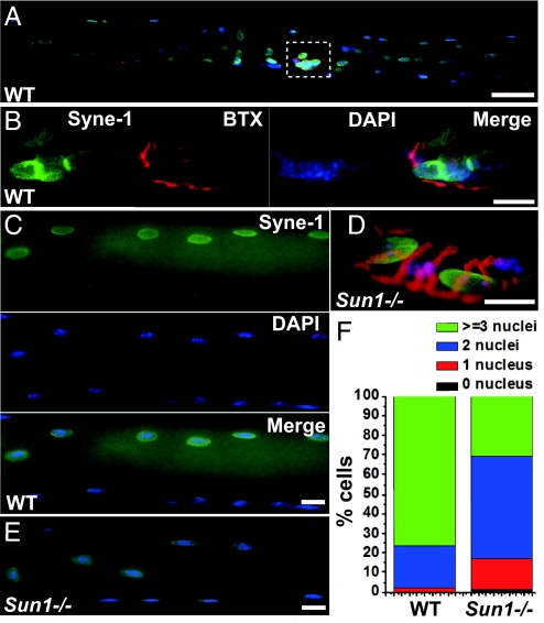
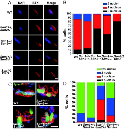
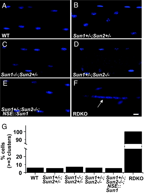
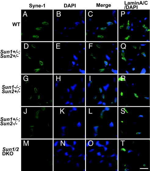
How the nuclei in mammalian skeletal muscle fibers properly position themselves relative to the cell body is an interesting and important cell biology question. In the syncytial skeletal muscle cells, more than 100 nuclei are evenly distributed at the periphery of each cell, with 3-8 nuclei anchored beneath the neuromuscular junction (NMJ). Our previous studies revealed that the KASH domain-containing Syne-1/Nesprin-1 protein plays an essential role in anchoring both synaptic and nonsynaptic myonuclei in mice. SUN domain-containing proteins (SUN proteins) have been shown to interact with KASH domain-containing proteins (KASH proteins) at the nuclear envelope (NE), but their roles in nuclear positioning in mice are unknown. Here we show that the synaptic nuclear anchorage is partially perturbed in Sun1, but not in Sun2, knockout mice. Disruption of 3 or all 4 Sun1/2 wild-type alleles revealed a gene dosage effect on synaptic nuclear anchorage. The organization of nonsynaptic nuclei is disrupted in Sun1/2 double-knockout (DKO) mice as well. We further show that the localization of Syne-1 to the NE of muscle cells is disrupted in Sun1/2 DKO mice. These results clearly indicate that SUN1 and SUN2 function critically in skeletal muscle cells for Syne-1 localization at the NE, which is essential for proper myonuclear positioning.


Conflict of interest statement

The authors declare no conflict of interest.

Figures

Fig. 1.
Fig. 1.
The anchorage of synaptic nuclei is partially perturbed in Sun1−/− mice. (A) A WT muscle fiber stained with anti–Syne-1 antibody (green), α-BTX (red), and DAPI (blue). (B) Enlarged view of the cropped region in (A), showing 4 synaptic nuclei clustered beneath a representative NMJ. (C) Part of the same image showing an even distribution of nonsynaptic nuclei along the periphery of the muscle fibers. [Scale bars: (A) 40 μm; (B and C) 10 μm.] (D and E) Representative images of synaptic nuclei (D) and nonsynaptic nuclei (E) in Sun1−/− mice. Only 2 nuclei are seen under the NMJ of this Sun1−/− muscle fiber. The distribution of nonsynaptic nuclei appears to be normal. (Scale bar: 10 μm.) (F) Statistical data showing that the average number of synaptic nuclei is decreased in Sun1−/− muscle fibers. In Sun1−/− mice, 15.9% of the fibers contain 1 synaptic nucleus, 52.3% contain 2 synaptic nuclei, and 30.7% contain 3 or more synaptic nuclei, compared with 1.8%, 21.4%, and 76.8% of respective WT fibers (WT, n = 56; Sun1−/−, n = 88; P < .001).
Fig. 2.
Fig. 2.
SUN1 and SUN2 play partially redundant roles in anchoring synaptic nuclei. (A) Fluorescent images showing skeletal muscle cells of triangularis sterni in the E18.5 embryos stained with DAPI (blue) and α-BTX (red). Genotypes of the embryos are indicated. (B) Statistical data showing the percentage of muscle fibers harboring various numbers of synaptic nuclei in mice with different genotypes. The respective percentages of muscle fibers harboring 0, 1, or 2 synaptic nuclei are 11%, 76%, and 13% in WT mice; 14%, 67%, and 19% in Sun1+/−; Sun2+/− mice; 63%, 36%, and 1% in Sun1−/−; Sun2+/− mice; 49%, 47%, and 4% in Sun1+/−; Sun2−/− mice; and 81%, 19%, and 0 in Sun1/2 DKO mice. Number of muscle cells scored (n): WT, n = 111; Sun1+/−; Sun2+/−, n = 122; Sun1−/−; Sun2+/−, n = 100; Sun1+/−; Sun2−/−, n = 122; Sun1/2 DKO, n = 107. (Scale bar: 5 μm.) (C) Representative images of synaptic myonuclei of the tibialis anterior muscle fibers in the adult mice of various genotypes stained with the anti–Syne-1 antibody (green), α-BTX (red), and DAPI (blue). (Scale bar: 10 μm.) (D) Statistical data showing the percentages of muscle fibers harboring various numbers of synaptic nuclei in mice with different genotypes. The respective percentages of muscle fibers harboring 0, 1, 2, or 3 or more synaptic nuclei are 0%, 1.8%, 21.4%, and 76.8% in WT mice (the same data as in Fig. 1F); 0%, 1.7%, 10.2%, and 88.1% in Sun1+/−; Sun2+/− mice; 6.1%, 51%, 34.7%, and 8.2% in Sun1−/−; Sun2+/− mice; and 1.6%, 15.6%, 43.8%, and 39% in Sun1+/−; Sun2−/− mice. Number of muscle cells scored: WT, n = 56; Sun1+/−; Sun2+/−, n = 59; Sun1−/−; Sun2+/−, n = 49; Sun1+/−; Sun2−/−, n = 64. These data indicate that both SUN1 and SUN2 contribute significantly to the anchorage of synaptic nuclei.
Fig. 3.
Fig. 3.
The organization of nonsynaptic nuclei is disrupted in −/−−/−RDKO mice. (A–F) The distribution of nonsynaptic nuclei in skeletal muscle fibers of adult mice stained with DAPI (blue). The arrow indicates a typical cluster of 6 nonsynaptic nuclei. (G) Statistical data showing the formation of myonuclear clusters in muscle fibers of −/−−/−RDKO mice. Number of muscle fibers scored: WT, n = 101; Sun1+/−; Sun2+/−, n = 104; Sun1−/−; Sun2+/−, n = 105; Sun1+/−; Sun2−/−, n = 109; Sun1+/−; Sun2−/−; NSE::Sun1: n = 102; RDKO, n = 50. (Scale bar: 10 μm.)
Fig. 4.
Fig. 4.
Expression of SUN1 in neurons rescues the lethality of Sun1/2 DKO mice. (A) The transgenic construct of the NSE-myc-Sun1 fusion gene. (B) Fluorescent images showing expression of the transgenic construct in the cortex of transgenic mice. The brain sections were stained with an anti–c-myc antibody (green), anti-SUN1 antibody (red), and DAPI (blue). (Scale bar: 10 μm.) (C) Fluorescent images showing no expression of the transgenic construct in skeletal muscle cells. (Scale bar: 10 μm.) (D) A RDKO mouse and its littermates at 8 days after birth. From left to right, the genotypes are Sun1+/−; Sun2−/−; NSE::Sun1, Sun1+/−; Sun2+/−, Sun1−/−; Sun2+/−; NSE::Sun1, and RDKO.
Fig. 5.
Fig. 5.
The NE localization of Syne-1 but not Lamin A/C is disrupted in Sun1/2 DKO mice. (A–O) Representative images of intercostal muscle sections of the E18.5 embryos stained with the anti–Syne-1 antibody (green) and DAPI (blue). Genotypes of the embryos are indicated. Syne-1 displays the NE localization pattern in muscle cells in WT (A–C), Sun1+/−; Sun2+/− (D–F), Sun1−/−; Sun2+/− (G–I), and Sun1+/−; Sun2−/− (J–L) mice, but not in the Sun1/2 DKO cells (M–O). (P–T) Representative merged images of intercostal muscle sections of the E18.5 embryos stained with the anti-Lamin A/C antibody (green) and DAPI (blue). Lamin A/C displays NE localization in muscle cells of all genotypes. (Scale bar: 10 μm.)

Similar articles

Cited by

References

    1. Sanes JR, Lichtman JW. Induction, assembly, maturation and maintenance of a postsynaptic apparatus. Nat Rev Neurosci. 2001;2:791–805. - PubMed
    1. Bruusgaard JC, Liestol K, Ekmark M, Kollstad K, Gundersen K. Number and spatial distribution of nuclei in the muscle fibres of normal mice studied in vivo. J Physiol. 2003;551(Pt 2):467–478. - PMC - PubMed
    1. Starr DA, Fischer JA. KASH 'n karry: The KASH domain family of cargo-specific cytoskeletal adaptor proteins. Bioessays. 2005;27:1136–1146. - PubMed
    1. Tzur YB, Wilson KL, Gruenbaum Y. SUN-domain proteins: “Velcro” that links the nucleoskeleton to the cytoskeleton. Nat Rev Mol Cell Biol. 2006;7:782–788. - PubMed
    1. Wilhelmsen K, Ketema M, Truong H, Sonnenberg A. KASH-domain proteins in nuclear migration, anchorage and other processes. J Cell Sci. 2006;119(Pt 24):5021–5029. - PubMed

Publication types

MeSH terms