. 2009 Jul;119(7):2042-51.
doi: 10.1172/JCI36131. Epub 2009 Jun 8.

Patients with lung cancer and paraneoplastic Hu syndrome harbor HuD-specific type 2 CD8+ T cells

Affiliations

Patients with lung cancer and paraneoplastic Hu syndrome harbor HuD-specific type 2 CD8+ T cells

Wendy K Roberts et al. J Clin Invest. 2009 Jul.

Abstract

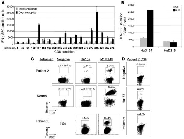
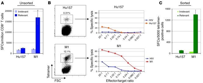
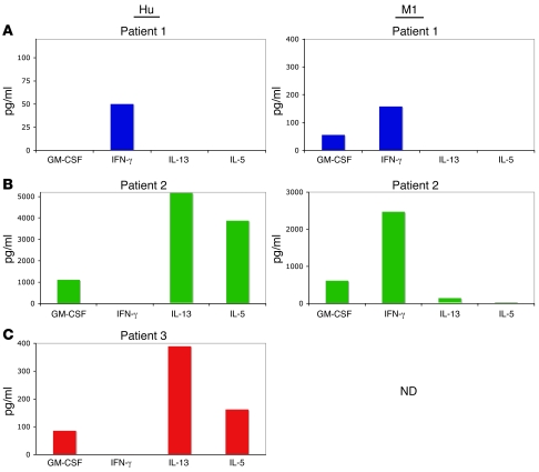
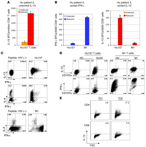
Paraneoplastic neurologic disorders (PNDs) offer an uncommon opportunity to study human tumor immunity and autoimmunity. In small cell lung cancer (SCLC), expression of the HuD neuronal antigen is thought to lead to immune recognition, suppression of tumor growth, and, in a subset of patients, triggering of the Hu paraneoplastic neurologic syndrome. Antigen-specific CTLs believed to contribute to disease pathophysiology were described 10 years ago in paraneoplastic cerebellar degeneration. Despite parallel efforts, similar cells have not been defined in Hu patients. Here, we have identified HuD-specific T cells in Hu patients and provided an explanation for why their detection has been elusive. Different Hu patients harbored 1 of 2 kinds of HuD-specific CD8+ T cells: classical IFN-gamma-producing CTLs or unusual T cells that produced type 2 cytokines, most prominently IL-13 and IL-5, and lacked cytolytic activity. Further, we found evidence that SCLC tumor cells produced type 2 cytokines and that these cytokines trigger naive CD8+ T cells to adopt the atypical type 2 phenotype. These observations demonstrate the presence of an unusual noncytotoxic CD8+ T cell in patients with the Hu paraneoplastic syndrome and suggest that SCLC may evade tumor immune surveillance by skewing tumor antigen-specific T cells to this unusual noncytolytic phenotype.


Figures

Figure 1
Figure 1. Demonstration of Hu133-specific T cells that have lytic activity and secrete IFN-γ.
(A) Tetramer staining of cells from patient 1 (HLA-0301) after 1 round of in vitro expansion with A0301-predicted peptides. The percentages of tetramer-positive cells are noted above the boxed populations. (B) The expanded T cells were also tested for functional activity in a CTL assay. Irrelevant peptide condition is designated by red squares, and relevant peptide is designated by blue circles. (C) Similar results were obtained with IFN-γ ELISPOT. Gray bars indicate irrelevant peptide condition, and black bars indicate relevant peptide. Error bars represent SD of the mean of triplicate wells (B and C).
Figure 2
Figure 2. Identification of the Hu157 epitope and Hu157-specific T cells in patients.
(A) Screening of Hu peptides in HLA-A0201 transgenic mice. CD8+ T cells purified from AAD mouse spleen cultures were cultured with stimulator cells pulsed with cognate or irrelevant peptide. A positive response was considered at least a 2-fold increase in signal over an irrelevant A0201 binding control peptide. (B) CD8+ T cells were tested for their ability to recognize whole HuD protein processed and presented by primary HHD kidney cells. Wild-type kidney cells were not recognized (data not shown). Peptide 315 was positive in the first screen (A) but was not processed on HLA-A0201 in the kidney cell experiment (B), indicating possible in vitro priming of T cells to this epitope or a low-affinity T cell response, and was not selected for further experiments with patient T cells. (C) Tetramer staining of patient 2, a normal donor, and patient 3 peripheral blood T cells after 1 round of in vitro expansion with HuD157 peptide. Positive control expansions and tetramer staining were with either M1 or CMV, depending on the response of the donor to these viruses. FSC, forward scatter; ND, not determined. (D) Tetramer staining for Hu157 T cells in CSF of Hu patient 2. The percentages of tetramer-positive cells are noted above the boxed populations in C and D. Error bars represent SD of the mean of triplicate wells (A and B).
Figure 3
Figure 3. Weak lytic activity and IFN-γ secretion by Hu157 patient T cells.
Hu157- and M1 tetramer–positive T cells from an Hu patient were expanded 1 time in vitro with peptide, followed by FACS sorting of the tetramer-positive population. Tetramer-positive cells were allowed to recover with irradiated, peptide-pulsed, autologous PBMCs and IL-2. After recovery, the function of the T cells was tested in a CTL assay. (A) ELISPOT with bulk (presort) CD8+ T cell cultures. (B) Tetramer staining showing sorting gate and CTL assay using sorted T cells. The percentages of tetramer-positive cells are noted above the boxed populations. (C) IFN-γ ELISPOT assay with sorted T cells. Error bars in A and C indicate SD of the mean of triplicate wells.
Figure 4
Figure 4. Screening for cytokines secreted by Hu-specific T cells reveals a Th2-like phenotype in some patients.
Supernatants from tetramer-sorted T cells cultured with target cells, pulsed with irrelevant or relevant peptide, were analyzed for the presence of 25 cytokines using a Multiplex kit. (A) Supernatants from patient 1, (B) from patient 2, and (C) from patient 3.
Figure 5
Figure 5. Hu patient T cells are skewed toward production of IL-13 and IL-4 but not IFN-γ.
(A) ELISPOT assay demonstrating the number of IL-13–producing, Hu-specific CD8+ cells in a bulk, unsorted Hu157 culture from patient 2 (same cells as in Figure 3A). (B) ELISPOT results using sorted cells from patient 3, comparing IFN-γ and IL-13 responses. Error bars in A and B indicate SD of the mean of triplicate wells. (C) Intracellular staining for IFN-γ, IL-13, and IL-4, using sorted T cells from Hu patient 2. The top 2 rows show Hu157 cells after irrelevant (HIV) and Hu157 peptide stimulation. The bottom row shows staining of M1-specific cells after irrelevant and M1 peptide stimulation. (D) Staining for Hu157- and M1-specific CD8+ T cells with lytic activity, using CD107a antibody combined with intracellular cytokine staining. The percentages of cells in each quadrant are denoted in the corners of the plots in C and D. (E) Intracellular staining of in vitro–skewed T cells showing production of IL-13 by CD8+ T cells and CD4+ T cells (positive controls) cultured in the presence of Th2 conditions (IL-4) but not Th1 conditions (IL-12, anti–IL-4). Numbers in corners represent the percentage of IL-13+ cells.

References

    1. Darnell R.B. Onconeural antigens and the paraneoplastic neurologic disorders: at the intersection of cancer, immunity and the brain. Proc. Natl. Acad. Sci. U. S. A. 1996;93:4529–4536. doi: 10.1073/pnas.93.10.4529. - DOI - PMC - PubMed
    1. Darnell R.B., Furneaux H.M., Posner J.B. Antiserum from a patient with cerebellar degeneration identifies a novel protein in Purkinje cells, cortical neurons, and neuroectodermal tumors. J. Neurosci. 1991;11:1224–1230. - PMC - PubMed
    1. Darnell R.B., Posner J.B. Paraneoplastic syndromes involving the nervous system. N. Engl. J. Med. 2003;349:1543–1554. doi: 10.1056/NEJMra023009. - DOI - PubMed
    1. Darnell R.B., Posner J.B. Paraneoplastic syndromes affecting the nervous system. Semin. Oncol. 2006;33:270–298. doi: 10.1053/j.seminoncol.2006.03.008. - DOI - PubMed
    1. Graus F., Elkon K.B., Cordon-Cardo C., Posner J.B. Sensory neuronopathy and small cell lung cancer; antineuronal antibody that also reacts with the tumor. Am. J. Med. 1986;80:45–52. doi: 10.1016/0002-9343(86)90047-1. - DOI - PubMed

Publication types