. 2009 Jun 9;4(6):e5846.
doi: 10.1371/journal.pone.0005846.

Aging and replicative senescence have related effects on human stem and progenitor cells

Affiliations

Aging and replicative senescence have related effects on human stem and progenitor cells

Wolfgang Wagner et al. PLoS One. .

Abstract

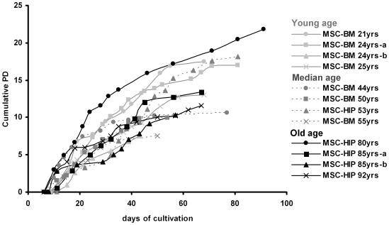
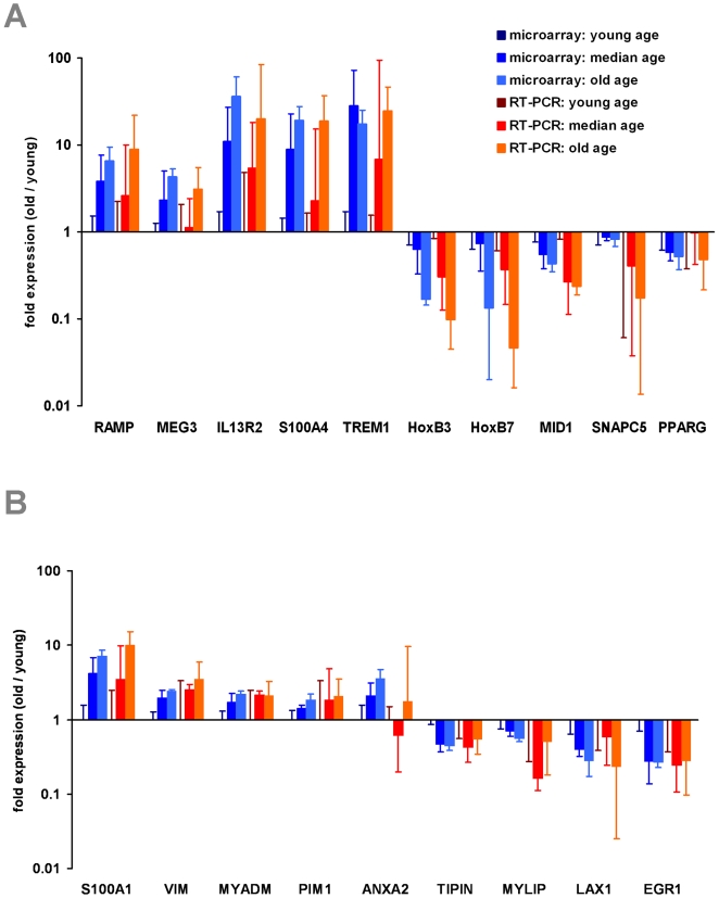
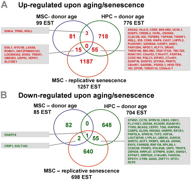
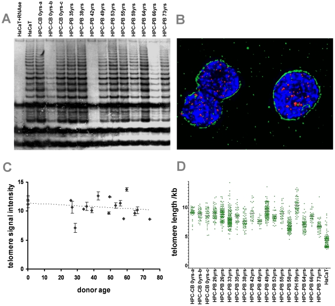
The regenerative potential diminishes with age and this has been ascribed to functional impairments of adult stem cells. Cells in culture undergo senescence after a certain number of cell divisions whereby the cells enlarge and finally stop proliferation. This observation of replicative senescence has been extrapolated to somatic stem cells in vivo and might reflect the aging process of the whole organism. In this study we have analyzed the effect of aging on gene expression profiles of human mesenchymal stromal cells (MSC) and human hematopoietic progenitor cells (HPC). MSC were isolated from bone marrow of donors between 21 and 92 years old. 67 genes were age-induced and 60 were age-repressed. HPC were isolated from cord blood or from mobilized peripheral blood of donors between 27 and 73 years and 432 genes were age-induced and 495 were age-repressed. The overlap of age-associated differential gene expression in HPC and MSC was moderate. However, it was striking that several age-related gene expression changes in both MSC and HPC were also differentially expressed upon replicative senescence of MSC in vitro. Especially genes involved in genomic integrity and regulation of transcription were age-repressed. Although telomerase activity and telomere length varied in HPC particularly from older donors, an age-dependent decline was not significant arguing against telomere exhaustion as being causal for the aging phenotype. These studies have demonstrated that aging causes gene expression changes in human MSC and HPC that vary between the two different cell types. Changes upon aging of MSC and HPC are related to those of replicative senescence of MSC in vitro and this indicates that our stem and progenitor cells undergo a similar process also in vivo.


Conflict of interest statement

Competing Interests: The authors have declared that no competing interests exist.

Figures

Figure 1
Figure 1. Long-term growth curves of mesenchymal stromal cells (MSC).
Cells were isolated from human bone marrow from the iliac crest (BM) or from the femoral head (HIP) of donors that were either young (21–25 years), median aged (44–55 years) or old (80–92 years). Cell numbers were determined at the end of every passage and cumulative population doublings (PD) were calculated in relation to the cell numbers at the first passage.
Figure 2
Figure 2. Age-induced gene expression in MSC is similar to replicative senescence.
Global gene expression profiles of 12 MSC samples of different donor age were analyzed by Affymetrix technology. Statistical analysis by template matching (PTM) revealed that 99 ESTs were significantly up-regulated (red) and 85 ESTs were down-regulated (green) with increasing donor age. These differentially expressed genes were further analyzed within the dataset of nine different passages of the same MSC donor sample (44 years old). Colour coding in the heat map demonstrates that gene expression changes upon aging are also reflected by replicative senescence of MSC in vitro.
Figure 3
Figure 3. QRT-PCR validation of differential gene expression.
Age associated gene expression changes in MSC were validated by quantitative RT-PCR. 10 genes were selected that have been differentially expressed in microarray data (A): regeneration associated muscle protease (RAMP); maternally expressed 3 (MEG3); interleukin 13 receptor, alpha 2 (IL13RA2); S100 calcium binding protein A4 (S100A4) and triggering receptor expressed on myeloid cells 1 (TREM1) were age-induced. Homeobox B3 (HOXB3) and Homeobox B7 (HOXB7); midline 1 (MID1); small nuclear RNA activating complex, polypeptide 5 (SNAPC5) and peroxisome proliferator-activated receptor gamma (PPARG) were age-repressed. Furthermore, we have validated age associated changes in HPC for 9 genes (B): S100 calcium binding protein A10 (S100A10); vimentin (VIM); myeloid-associated differentiation marker (MYADM); pim-1 oncogene (PIM1) and annexin A2 (ANXA2) were age-induced. Timeless interacting protein (TIPIN); myosin regulatory light chain interacting protein (MYLIP); lymphocyte transmembrane adaptor 1 (LAX1) and Early growth response 1 (ERG1) were age-repressed. Protocadherin 9 (PCDH9) was not amplified in HPC from elderly donors whereas interleukine 7 receptor (IL7R) was not amplified in young samples (not presented in the figure). Differential gene expression was always calculated in relation to the mean of young samples. The mean fold-ratio (±SD) is demonstrated for median aged and old donor samples. RT-PCR results (red) were always in line with microarray data (blue) for all genes tested.
Figure 4
Figure 4. Age-induced gene expression of hematopoietic progenitor cells (HPC).
CD34+ HPC were isolated from cord blood (CB) or from mobilized peripheral blood (PB) of healthy donors (27–73 years). Gene expression profiles revealed that 776 ESTs were significantly age-induced (red) and 704 ESTs were age-repressed (green). We have combined the age-induced gene expression of HPC with replicative senescence data in MSC. The heat map demonstrated here indicates an association between the two data sets.
Figure 5
Figure 5. Combination of the three datasets.
Differential gene expression of the three datasets (MSC–donor age; HPC–donor age and MSC–replicative senscence) was further compared. Venn diagrams demonstrate the overlap of differentially expressed ESTs that are up-regulated upon aging (A) or down-regulated (B). Differentially expressed genes are indicated as short cut (Hugo name). There was only little overlap between MSC–donor age and HPC–donor age. However, both datasets demonstrated a relatively high overlap with the dataset MSC–replicative senescence.
Figure 6
Figure 6. Telomerase activity and 3D reconstruction of telomeres in HPC upon aging.
TRAP assay was performed on CD34+ cells of different donor ages. HaCaT cells exhibiting a similar telomerase activity as HeLa cells were used as a positive control . Telomerase activity in HPC varied between hardly detectable and as high as in HaCaT cells with no clear association between the level of telomerase activity and donor age (A; HaCaT cell lysat = positive control, HaCaT lysat + RNAse = negative control). Telomere length in HPC was estimated using 3D image reconstruction. Cells were immunostained for CD34 (green), hybridized for telomeres with the telomere-specific PNA probe (red) and DNA counterstained with DAPI (blue) (B). Graphic presentation of the telomere signal intensity measurement (arbitrary units) of CD34+ cells from 19 different age donors demonstrates a weak tendency of telomere shortening during age (C; the slope of linear regression is not significant, P = 0.1034). The distribution of telomere length in individual cells revealed a tendency for higher variation in telomere length in HPC of elderly donors (D; P = 0.0450).

References

    1. Ho AD, Wagner W, Mahlknecht U. Stem cells and ageing. The potential of stem cells to overcome age-related deteriorations of the body in regenerative medicine. EMBO Rep. 2005;6:35–38. - PMC - PubMed
    1. Gazit R, Weissman IL, Rossi DJ. Hematopoietic stem cells and the aging hematopoietic system. Semin Hematol. 2008;45:218–224. - PubMed
    1. Giangreco A, Qin M, Pintar JE, Watt FM. Epidermal stem cells are retained in vivo throughout skin aging. Aging Cell. 2008;7:250–259. - PMC - PubMed
    1. Horwitz EM, Le BK, Dominici M, Mueller I, Slaper-Cortenbach I, et al. Clarification of the nomenclature for MSC: The International Society for Cellular Therapy position statement. Cytotherapy. 2005;7:393–395. - PubMed
    1. Horn P, Bork S, Diehlmann A, Walenda T, Eckstein V, et al. Isolation of human mesenchymal stromal cells is more efficient by red blood cell lysis. Cytotherapy. 2008;10:676–685. - PubMed

Publication types