. 2009 Jun 9;4(6):e5839.
doi: 10.1371/journal.pone.0005839.

SHMT1 and SHMT2 are functionally redundant in nuclear de novo thymidylate biosynthesis

Affiliations

SHMT1 and SHMT2 are functionally redundant in nuclear de novo thymidylate biosynthesis

Donald D Anderson et al. PLoS One. .

Abstract

The three enzymes that constitute the de novo thymidylate synthesis pathway in mammals, cytoplasmic serine hydroxymethyltransferase (SHMT1), thymidylate synthase (TYMS) and dihydrofolate reductase (DHFR) undergo sumoylation and nuclear import during S-phase. In this study, we demonstrate that purified intact mouse liver nuclei convert dUMP to dTMP in the presence of NADPH and serine. Neither nuclear extracts nor intact nuclei exposed to aminomethylphosphonate, a SHMT inhibitor, exhibit thymidylate synthesis activity. Nuclei isolated from Shmt1(-/-) mouse livers retained 25% of thymidylate synthesis activity exhibited by nuclei isolated from wild type mice. This residual activity was due to the presence of a cytoplasmic/nuclear isozyme of SHMT encoded by Shmt2. Shmt2 is shown to encode two transcripts, one which encodes a protein that localizes exclusively to the mitochondria (SHMT2), and a second transcript that lacks exon 1 and encodes a protein that localizes to the cytoplasm and nucleus during S-phase (SHMT2alpha). The ability of Shmt2 to encode a cytoplasmic isozyme of SHMT may account for the viability of Shmt1(-/-) mice and provide redundancy that permitted the expansion of the human SHMT1 L474F polymorphism that impairs SHMT1 sumoylation and nuclear translocation.


Conflict of interest statement

Competing Interests: The authors have declared that no competing interests exist.

Figures

Figure 1
Figure 1. Compartmentation of folate-mediated one-carbon metabolism in the cytoplasm, mitochondria and nucleus.
One-carbon metabolism in the cytoplasm is required for the de novo synthesis of purines and thymidylate, and for the remethylation of homocysteine to methionine. One-carbon metabolism in mitochondria generates one-carbon units for cytoplasmic one-carbon metabolism by generating formate from serine and glycine. One-carbon metabolism in the nucleus synthesizes dTMP from dUMP and serine. SHMT2, mitochondrial serine hydroxymethyltransferase; SHMT1, cytoplasmic serine hydroxymethyltransferase; TYMS, thymidylate synthase; DHFR, dihydrofolate reductase; THF, tetrahydrofolate.
Figure 2
Figure 2. Thymidylate biosynthesis occurs in purified nuclei.
Liver nuclei were isolated from SHMT1+/+ and SHMT1−/− mice and capacity to convert dUMP and [2,3-3H]-L-serine to [3H]dTMP was determined in reactions that contained: 1) sonicated nuclei; 2) intact nuclei; 3) intact nuclei with 200 µM 5-CHOTHFGlu5; and 4) intact nuclei with 100 mM aminomethyl phosphonate (AMPA). De novo thymidylate biosynthesis activities were normalized to activity generated from SHMT1+/+ intact nuclei which was given an arbitrary value of 1.0. Reactions containing sonicated nuclei contained no activity. All reactions were performed in duplicate and the experiment repeated twice. Variation is expressed as the standard deviation.
Figure 3
Figure 3. Identification of SHMT2 in purified nuclei.
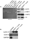
Panel A) PCR was used to confirm the genotype of nuclei isolated from SHMT1+/+ and SHMT1−/− mice. SHMT1−/− mice were generated through deletion of exon 7 which encodes the PLP binding site. SHMT1+/+ mice exhibit a 740-bp PCR product whereas the SHMT1−/− mice exhibit a 460-bp PCR product. Western blotting confirmed the presence of TYMS and DHFR in nuclei of both genotypes. SHMT1 was present in purified liver nuclei from SHMT1+/+ mice, but absent in liver nuclei from SHMT1−/− mice. SHMT2 was present in nuclei from both genotypes. Panel B) Western blots confirm the purity of the isolated liver nuclei. The control lane represents purified cytosol from NIH/3T3 cells for both the Lamin A (nuclear marker) and GAPDH (cytoplasmic marker). The control for the COX IV immunoblot represents a purified mitochondrial fraction from NIH/3T3 cells. The absence of GAPDH and COX IV in nuclear extracts indicated that no cytosolic or mitochondrial contamination was present in the nuclear thymidylate biosynthetic assays.
Figure 4
Figure 4. The mouse Shmt2 encodes two transcripts and contains two translation initiation sites.
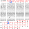
The SHMT2 transcript is denoted by the color red. The first nucleotide of the translation initiation site contained in exon 1 is numbered as +1. The first translation initiation site in exon 1, which encodes for the mitochondrial leader sequence, is boxed in blue. The mitochondrial leader sequence is cleaved co-translationally in the mitochondria denoted by the scissors . The second translation initiation site, present in exon 2, is boxed in blue. A transcription start site in intron 1 deduced from a mouse liver EST (AA793217) is denoted by the arrow and starts at −166 from the second ORF.
Figure 5
Figure 5. SHMT2α localizes to the cytoplasm and nucleus.
HeLa cells were transfected with cDNAs encoding SHMT2-YFP, mito-CFP, and SHMT2α-RFP. A) The SHMT2-YFP fusion protein expressed in HeLa cells localizes to mitochondria. The mitochondrial marker, mito-CFP has a similar localization pattern as that of SHMT2. The SHMT2α-RFP fusion protein expressed in HeLa cells localizes to both the cytoplasm and the nucleus. B) SHMT2α-RFP localizes to the nucleus in a cell cycle dependent manner during S-phase and G2/M, whereas it is absent from the nucleus in G1 phase.
Figure 6
Figure 6. SHMT2 and SHMT2α rescue the glycine auxotrophy in CHO glyA cells.
CHO glyA cells were transfected with cDNAs encoding human SHMT2-RFP, SHMT2α−RFP, and a RFP-empty vector control. Stable cells lines were selected for G418 resistance in the presence of 200 mM glycine. For growth assays, cells were cultured with and without glycine and MTT assays were used to quantify growth. Twelve independent lines were assayed per transfection and experiments were done in triplicate. All values are normalized to RFP-empty vector transfectants. There was no significant difference in growth among the cells transfected with SHMT2 and SHMT2α with or without glycine. RFP-empty vector transfectants with and without glycine are shown as a circle and open box respectively. SHMT2-RFP transfectants with and without glycine are shown as a closed box and triangle respectively. SHMT2α−RFP transfectants with and without glycine are shown as an inverted triangle and diamond respectively.
Figure 7
Figure 7. SHMT2 and SHMT2α are present in mouse tissues.
Immunoblots using a sheep-anti-human SHMT2 antibody revealed the presence of three immunoreactive bands. Protein masses were determined by the migration distance and relative mobility of standards. Purified liver nuclei, whole liver and whole kidney extracts contained an immunoreactive band at ∼53 kDa, the predicted mass of SHMT2α. Purified liver mitochondria, whole liver and whole kidney extracts contained a band at ∼56 kDa, the predicted mass of the SHMT2 pre-processed protein. Purified liver mitochondria and whole liver, but not whole kidney extracts contained a band at ∼50 kDa, the predicted mass of the SHMT2 processed protein. The nuclear fraction was free of cytosolic and mitochondrial contamination as shown by the GAPDH and COX IV immunoblots.
Figure 8
Figure 8. The L474F polymorphism in SHMT1 impairs nuclear localization.
SHMT1−/− MEF cells were transfected with cDNAs encoding the YFP-empty vector, YFP-SHMT1 wild-type, YFP-SHMT1-K38R/K39R, and YFP-SHMT1-L474F. S-phase blocked transfectants showed that YFP-SHMT1 is greatly increased in the nucleus in S-phase. Empty vector control and K38R/K39R mutation eliminated nuclear localization, and the L474F polymorphism inhibited nuclear localization.

References

    1. Fox JT, Stover PJ. Folate-mediated one-carbon metabolism. Vitam Horm. 2008;79:1–44. - PubMed
    1. Garrow TA, Brenner AA, Whitehead VM, Chen XN, Duncan RG, et al. Cloning of human cDNAs encoding mitochondrial and cytosolic serine hydroxymethyltransferases and chromosomal localization. J Biol Chem. 1993;268:11910–11916. - PubMed
    1. Girgis S, Nasrallah IM, Suh JR, Oppenheim E, Zanetti KA, et al. Molecular cloning, characterization and alternative splicing of the human cytoplasmic serine hydroxymethyltransferase gene. Gene. 1998;210:315–324. - PubMed
    1. Stover PJ, Chen LH, Suh JR, Stover DM, Keyomarsi K, et al. Molecular cloning, characterization, and regulation of the human mitochondrial serine hydroxymethyltransferase gene. J Biol Chem. 1997;272:1842–1848. - PubMed
    1. Appling DR. Compartmentation of folate-mediated one-carbon metabolism in eukaryotes. Faseb J. 1991;5:2645–2651. - PubMed

Publication types