. 2009 Jun 9;4(6):e5822.
doi: 10.1371/journal.pone.0005822.

Modulation of eDNA release and degradation affects Staphylococcus aureus biofilm maturation

Affiliations

Modulation of eDNA release and degradation affects Staphylococcus aureus biofilm maturation

Ethan E Mann et al. PLoS One. .

Abstract

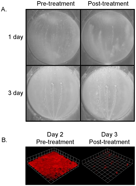
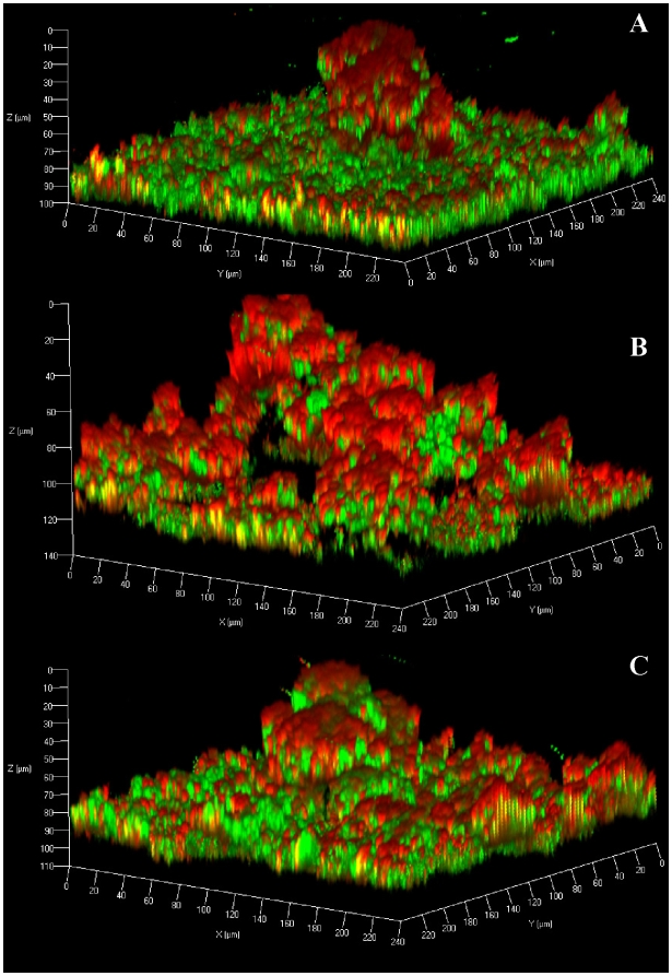
Recent studies have demonstrated a role for Staphylococcus aureus cidA-mediated cell lysis and genomic DNA release in biofilm adherence. The current study extends these findings by examining both temporal and additional genetic factors involved in the control of genomic DNA release and degradation during biofilm maturation. Cell lysis and DNA release were found to be critical for biofilm attachment during the initial stages of development and the released DNA (eDNA) remained an important matrix component during biofilm maturation. This study also revealed that an lrgAB mutant exhibits increased biofilm adherence and matrix-associated eDNA consistent with its proposed role as an inhibitor of cidA-mediated lysis. In flow-cell assays, both cid and lrg mutations had dramatic effects on biofilm maturation and tower formation. Finally, staphylococcal thermonuclease was shown to be involved in biofilm development as a nuc mutant formed a thicker biofilm containing increased levels of matrix-associated eDNA. Together, these findings suggest a model in which the opposing activities of the cid and lrg gene products control cell lysis and genomic DNA release during biofilm development, while staphylococcal thermonuclease functions to degrade the eDNA, possibly as a means to promote biofilm dispersal.


Conflict of interest statement

Competing Interests: The authors have declared that no competing interests exist.

Figures

Figure 1
Figure 1. Extracellular DNA-mediated attachment of static biofilm.
S. aureus UAMS-1 static biofilms were treated with either DNase I (grey bars) or PAS (black bars) at the time of inoculation (t = 0), and at 2, 4, 6, 10, and 24 hours post-inoculation. All biofilms were grown at 37°C for a total of 24 hours. 24-hour biofilms were allowed to grow for several hours after PAS or DNase I addition, to allow full penetration and activity of the compound on the biofilm. The biofilms were washed, stained with crystal violet, and retained biomass was quantified by measuring the absorbance of each well at an absorbance of 655 nm. Mean values from three independent experiments are shown and error bars represent the SEM.
Figure 2
Figure 2. Static assays and eDNA quantification.
S. aureus static biofilms were grown for 24 hours. (A) Washed biofilms were stained with crystal violet and quantified spectrophotometrically at an absorbance of 595 nm. The decrease in the cidA mutant (KB1050) biofilm adherence observed compared to the wild-type strain (UAMS-1) was statistically significant (p = 0.0004; ANOVA), as was the increased lrgAB mutant (KB1045) biofilm adherence (p = 0.021). There is no significant difference between the complementation strain (KB1046) (p = 0.39) and the UAMS-1 phenotype. (B) Extracellular DNA was isolated from the biofilm matrices of UAMS-1, KB1050, KB1045, and KB1046 and qRT-PCR of four chromosomal loci were amplified, gyr (black bars), lue (slashed bars), lys (grey bars), and fhu (white bars). The relative biomass was quantified at OD600, and the eDNA measurements were normalized to total biofilm biomass as described previously . The relative concentration of eDNA decreases in the cidA mutant (p<0.0001) and increases in the in the lrgAB mutant (p = 0.020) compared to the wild-type. There is no significant difference between the complementation strain (KB1046) (p = 0.36) and the wild-type. Results are depicted as averages of three independent experiments and error bars represent the SEM.
Figure 3
Figure 3. Flow cell biofilm assays.
S. aureus 3-day biofilms were grown and representative images were taken using a macroscopic camera (panels A, C, E, and G) or CLSM (panels B, D, F, and H). The samples were stained with Syto-9 (green) and propidium iodide (PI; red) to indicate live and dead cell populations, respectively. The UAMS-1 (A and B), cidA mutant (C and D), lrgAB mutant (E and F), and lrgAB complemented (G and H) biofilms were grown for three days in the flow-cell model prior to imaging. The images shown are representative of four independent experiments.
Figure 4
Figure 4. Visualisation of eDNA.
S. aureus flow-cell biofilms were stained with Syto-9 and Toto-3 to indicate the location of live cells (green) and dead cells (punctuate red) and extracellular DNA (diffuse red). A yellow appearance is observed where live cells and eDNA are both present. The UAMS-1 (A), cidA mutant (B), and lrgAB mutant (C) biofilms were grown for three days prior to imaging and representative CLSM images are shown. The images shown are representative of three independent experiments.
Figure 5
Figure 5. Mature biofilm sensitivity to DNase I.
(A) S. aureus UAMS-1 biofilms were grown in the flow-cell model with low inoculum for one or three days. The images of untreated biofilms where taken immediately before DNase I exposure and the images of the same biofilms treated with DNase I were taken five hours later. (B) S. aureus UAMS-1 biofilms harboring a constitutively-expressed gene (rfp) encoding red fluorescence protein were grown in the same flow-cell model using a higher inoculum for two days. The biofilm was then treated with DNase I for a total of 24 hours after which CLSM z-stacks were taken. Renderings of the z-stack CLSM data were performed using the Volocity software.
Figure 6
Figure 6. Staphylococcal thermonuclease affects biofilm structure.
Representative CLSM z-stacks of UAMS-1 (A), nuc mutant (B), and nuc complemented (C) biofilms stained with Syto-9 (green) and Toto-3 (red). Syto-9 represents the live cells and Toto-3 represents dead cells and eDNA. The images shown are representative of three independent experiments.

References

    1. Costerton JW, Cheng KJ, Geesey GG, Ladd TI, Nickel JC, et al. Bacterial biofilms in nature and disease. Annu Rev Microbiol. 1987;41:435–464. - PubMed
    1. Costerton JW, Lewandowski Z, DeBeer D, Caldwell D, Korber D, et al. Biofilms, the customized microniche. J Bacteriol. 1994;176:2137–2142. - PMC - PubMed
    1. Boles BR, Horswill AR. Agr-mediated dispersal of Staphylococcus aureus biofilms. PLoS Pathog. 2008;4:e1000052. - PMC - PubMed
    1. Vuong C, Otto M. The biofilm exopolysaccharide polysaccharide intercellular adhesion–a molecular and biochemical approach. Methods Mol Biol. 2008;431:97–106. - PubMed
    1. Otto M. Staphylococcal biofilms. Curr Top Microbiol Immunol. 2008;322:207–228. - PMC - PubMed

Publication types

LinkOut - more resources