

Intracranial hemodynamic changes during adult moyamoya disease progression
- PMID: 19513306
- PMCID: PMC2686870
- DOI: 10.3988/jcn.2008.4.2.67
Intracranial hemodynamic changes during adult moyamoya disease progression
Abstract
Background and purpose: This study evaluated the changes in blood flow velocity in the anterior and posterior intracranial circulations according to the progression of moyamoya disease in adult patients.
Methods: We evaluated Suzuki's angiographic stage and mean blood flow velocity (MBFV) changes in intracranial vessels from both sides in 19 adult moyamoya patients. We then analyzed the linearity of MBFV changes from early to late moyamoya stages in each intracranial vessel using piecewise linear regression models.
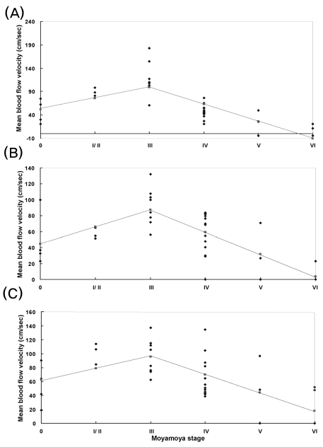
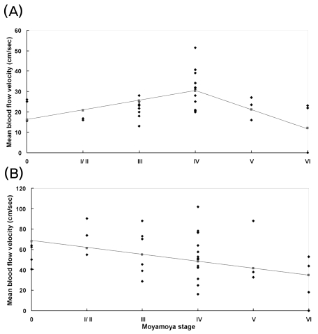
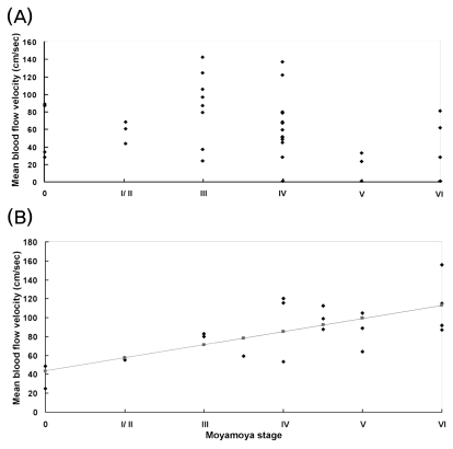
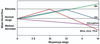
Results: The MBFV in the middle cerebral artery, terminal internal carotid artery, and anterior cerebral artery increased non linearly until stage III, and then decreased progressively up to stage VI. The ophthalmic artery also showed nonlinear velocity changes, with an increase in MBFV up to stage IV, followed by a decrease in MBFV up to stage VI. The MBFV of the basilar artery increased linearly from a normal velocity at an early moyamoya stage to a stenotic velocity at a late stage. There was no statistically significant regression model for the relationship between the MBFV in the posterior cerebral artery and moyamoya stage.
Conclusions: The nonlinear and/or linear MBFV changes associated with variable intracranial vessels might be useful in initial and follow-up evaluations of different stages of moyamoya disease.
Keywords: Cerebral blood flow; Moyamoya disease; Transcranial Doppler.
Figures
References
-
- Suzuki J, Takaku A. Cerebrovascular "moyamoya" disease. Disease showing abnormal net-like vessels in base of brain. Arch Neurol. 1969;20:288–299. - PubMed
-
- Takase K, Kashihara M, Hashimoto T. Transcranial Doppler ultrasonography in patients with moyamoya disease. Clin Neurol Neurosurg. 1997;99(Suppl 2):S101–S105. - PubMed
-
- Lee YS, Jung KH, Roh JK. Diagnosis of moyamoya disease with transcranial Doppler sonography: correlation study with magnetic resonance angiography. J Neuroimaging. 2004;14:319–323. - PubMed
-
- Yamauchi T, Tada M, Houkin K, Tanaka T, Nakamura Y, Kuroda S, et al. Linkage of familial moyamoya disease (spontaneous occlusion of the circle of Willis) to chromosome 17q25. Stroke. 2000;31:930–935. - PubMed
-
- Suzuki J. Moyamoya Disease. Berlin: Springer-Verlag; 1983. pp. 143–185.
LinkOut - more resources
Full Text Sources
