. 2009 Dec;19(12):2941-9.
doi: 10.1007/s00330-009-1474-7.

Congenital heart diseases: post-operative appearance on multi-detector CT-a pictorial essay

Affiliations

Congenital heart diseases: post-operative appearance on multi-detector CT-a pictorial essay

Anderanik Tomasian et al. Eur Radiol. 2009 Dec.

Abstract

Echocardiography is considered as an initial imaging modality of choice in patients with congenital heart disease (CHD), and magnetic resonance (MR) imaging is preferred for detailed functional information. Multi-detector computed tomography (CT) plays an important role in clinical practice in assessing post-operative morphological and functional information of patients with complex CHD when echocardiography and MR imaging are not contributory. Radiologists should understand and become familiar with the complex morphology and physiology of CHD, as well as with various palliative and corrective surgical procedures performed in these patients, to obtain CT angiograms with diagnostic quality and promptly recognise imaging features of normal post-operative anatomy and complications of these complex surgeries.


Figures

Fig. 1
Fig. 1
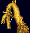
Blalock–Taussig shunt. a Volume-rendered image (posterior view) in a 43-year-old man with a history of ToF demonstrates a patent, classic, right BT shunt with mild distal narrowing (arrow). b Oblique coronal MIP image in a different patient with ToF shows stenosis of the left classic BT shunt (arrow). In a modified BT shunt where the connection between the subclavian and ipsilateral pulmonary artery is created by a graft, blood supply to the ipsilateral upper extremity is preserved via the subclavian artery
Fig. 2
Fig. 2
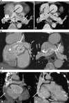
Davidson (Central) shunt. a Oblique coronal MIP image in a 16-year-old girl with hypoplastic left heart syndrome and status post-Davidson (Central) shunt demonstrates a patent Gore-Tex shunt graft connecting the ascending aorta to the main pulmonary artery (arrow). b Axial MIP image in a 14-year-old boy with a history of TGA, a single AV canal and subaortic stenosis shows a thrombosed Davidson (Central) shunt (arrow)
Fig. 3
Fig. 3
Waterston–Cooley shunt. Volume-rendered image in a 14-year-old boy with a history of ToF and pulmonary atresia demonstrates a Waterston–Cooley shunt extending from the posterior wall of the ascending aorta and the anterior wall of the right pulmonary artery. Note the mild narrowing in the mid portion of the shunt (arrow)
Fig. 4
Fig. 4
Damus–Kaye–Stansel shunt. Oblique coronal MIP image in a 14-year-old boy with a history of TGA shows the transected main pulmonary artery with connection to the ascending aorta by an end-to-side anastomosis (large arrow). Note the extra-cardiac Fontan shunt (small arrow; not opacified). A later phase image (not shown) demonstrated contrast opacification of the Fontan without thrombosis
Fig. 5
Fig. 5
Norwood–Sano procedure. Oblique sagittal MIP images in a 20-year-old woman with a history of hypoplastic left heart syndrome demonstrate ascending aortic reconstruction using the proximal main pulmonary artery (a, large arrow). The main pulmonary artery continues as the aortic arch and descending aorta (a, small arrow). Hypoplastic conventional ascending aorta arises from the left ventricle (b, large arrow). The conventional origin of the right coronary artery is also noted (b, small arrow)
Fig. 6
Fig. 6
a Bidirectional Glenn shunt with a left pulmonary artery stent. Oblique coronal and axial MIP images in a 19-year-old man with tricuspid atresia and an atrioventricular canal defect demonstrate anastomosis of the SVC to the right main pulmonary artery (A and B, large arrow). Noted is a left main pulmonary artery stent without in-stent re-stenosis (A and B, small arrow). b Bidirectional Glenn shunt. Axial MIP image in a 24-year-old man with a history of TGA demonstrates anastomosis of the SVC to the main pulmonary artery confluence (arrow)
Fig. 7
Fig. 7
a Direct classic Fontan shunt. Axial MIP images in a 26-year-old man with a history of ToF demonstrate direct anastomosis of the RA to the main PA confluence (A and B, black arrow). In the early phase (A), there is a thrombosis-like appearance of bilateral PAs because of mixing artefact (A, white arrows). In the later phase (B), thromboemboli are clearly seen in the RA and the right PA (B, small black arrows) with good opacification and absent mixing artefact of the main PAs. b Modified Fontan shunt. Oblique coronal MIP image in a 35-year-old man with a history of TGA and tricuspid atresia demonstrates the connection between the right atrium (A, arrowhead) and the main PA confluence through a Gore Tex graft (A, arrow) that is peripherally calcified. Noted is a left main PA stent (B, arrow). c Total cavopulmonary connection with extra-cardiac Fontan shunt. Oblique coronal MIP images in a 25-year-old man with a history of TGA demonstrate the anastomosis of the IVC (A and B, large arrow) to the main PA confluence (A and B, small arrow). In the arterial phase (A) the Fontan shunt is not opacified, resembling a thrombosis. In the venous phase (B), normal opacification of the shunt is noted, indicating patency. The patient also has a bidirectional superior vena cava (SVC) Glenn shunt. In this procedure, the SVC flow is directed mostly to the right pulmonary artery, and the IVC flow to the left PA
Fig. 8
Fig. 8
Rastelli procedure. Oblique sagittal MIP images in a 30-year-old woman with a history of complex CHD demonstrate the connection of the pulmonary artery confluence to the right ventricle with an extra-cardiac, peripherally calcified conduit without significant stenosis (a and b, large arrow). Note the atretic main pulmonary artery (b, arrowhead). The VSD is closed by a Gore Tex graft (a, small arrow) and the LV is connected to the aorta. The right coronary artery is also noted to originate from the aorta (a, arrowhead)
Fig. 9
Fig. 9
Pulmonary artery banding. Axial MIP image in a 15-year-old girl with a history of hypoplastic left heart syndrome shows proximal right pulmonary artery banding (arrow)
Fig. 10
Fig. 10
Unifocalisation. Oblique coronal MIP image in a 14-year-old girl with a history of ToF with pulmonary atresia and major aortopulmonary collaterals shows right unifocalisation (large arrow) with Gore Tex graft extension (small arrow) into left unifocalisation (arrowhead) and the right ventricular outflow tract (black arrow)
Fig. 11
Fig. 11
Atrial switch repair. A 19-year-old man’s status post-Mustard procedure for double outlet right ventricle and TGA. Oblique axial MIP image demonstrates an inter- atrial baffle (a, large arrow) separating the pulmonary venous chamber and the systemic venous chamber. The main pulmonary artery arises from the double-outlet right ventricle (a, small arrow). Oblique coronal MIP image demonstrates the pulmonary venous chamber (b, arrow) and drainage of the IVC and SVC to the systemic venous chamber (b, arrowhead)
Fig. 12
Fig. 12
a Jatene procedure (arterial switch repair). An 18-year-old man with a history of D-TGA and status post-Jatene procedure. Oblique coronal and axial MIP images demonstrate switching of the aorta (A and B, large arrow) and pulmonary artery (A and B, small arrows) along with the transposition of the coronary arteries (C, arrows) from the aorta (or neo-pulmonary artery) to the pulmonary artery (or neo-aorta). Note that the neopulmonary artery is located anterior to the neo-aorta (Lecompte manoeuvre). b Jatene procedure (arterial switch repair).Volume-rendered (A) and oblique axial MIP image (B) in a 22-year-old man with a history of D-TGA and status post-Jatene procedure. Note the left pulmonary artery stenosis (A and B, large arrow) by the neo-aorta (A and B, small arrow)

Similar articles

Cited by

References

    1. Haramati N, Crooke GA. MR imaging and CT of vascular anomalies and connections in patients with congenital heart disease: significance in surgical planning. Radiographics. 2002;22:337–347. - PubMed
    1. Masui T, Katayama M, Kobayashi S, et al. Gadolinium-enhanced MR angiography in the evaluation of congenital cardiovascular disease pre- and postoperative states in infants and children. J Magn Reson Imaging. 2000;12:1034–1042. doi: 10.1002/1522-2586(200012)12:6<1034::AID-JMRI32>3.0.CO;2-A. - DOI - PubMed
    1. Flohr T, Stierstorfer K, Raupach R, Ulzheimer S, Bruder H. Performance evaluation of a 64-slice CT system with z-flying focal spot. Rofo. 2004;176:1803–1810. - PubMed
    1. Alkhulaifi AM, Lacour-Gayet F, Serraf A, Belli E, Planche C. Systemic pulmonary shunts in neonates: early clinical outcome and choice of surgical approach. Ann Thorac Surg. 2000;69:1499–1504. doi: 10.1016/S0003-4975(00)01078-X. - DOI - PubMed
    1. Rodriguez E, Soler R, Rernandez R, Raposo I. Postoperative imaging in cyanotic congenital heart diseases: part I, normal findings. AJR Am J Roentgenol. 2007;189:1353–1360. doi: 10.2214/07.2104. - DOI - PubMed