Review
. 2009 Aug;21(4):522-30.
doi: 10.1016/j.ceb.2009.04.015. Epub 2009 Jun 8.

Turnover of organelles by autophagy in yeast

Affiliations
Review

Turnover of organelles by autophagy in yeast

Jean-Claude Farré et al. Curr Opin Cell Biol. 2009 Aug.

Abstract

Efficient detection and removal of superfluous or damaged organelles are crucial to maintain cellular homeostasis and to assure cell survival. Growing evidence shows that organelles or parts of them can be removed by selective subtypes of otherwise unselective macroautophagy and microautophagy. This requires both the adaptation of the core autophagic machinery and sophisticated mechanisms to recognize organelles destined for turnover. We review the current knowledge on autophagic removal of peroxisomes, mitochondria, ER and parts of the nucleus with an emphasis on yeasts as a model eukaryote.


Figures

Figure 1
Figure 1. Morphology of macroautophagy and the Cvt pathway in S. cerevisiae.
Starvation-induced macroautophagy starts with the formation of a double-layered isolation membrane or phagophore (nucleation). After elongation of this structure, fusion of the edges yields an autophagosome. The fusion of the autophagosomal outer membrane with the vacuole eventually releases an autophagic body into the vacuole for degradation. The morphologically similar Cvt pathway uses smaller transport vesicles, whose biogenesis starts at a cargo complex containing pApeI.
Figure 2
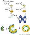
Figure 2. Scheme of the two ubiquitin-like conjugation systems involved in macroautophagy
After proteolytic processing of its carboxyterminus by the Atg4 protease, Atg8 is coupled to the membrane lipid phosphatidylethanolamine (PE). A second conjugation system attaches Atg12 to Atg5. The Atg12-Atg5 conjugate then forms an oligomer with Atg16 and stimulates the formation of Atg8-PE. As indicated, after elongation of the phagophore the Atg12-Atg5/Atg16 oligomer predominantly localizes to the outer membrane. The release of Atg8 from the mature autophagosome requires Atg4. Green circles: Atg8-PE.
Figure 3
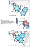
Figure 3. Macro- and micropexophagy model
A late stage of pexophagy is represented in the figure, which shows the PAS, PC, PVS, VSM, MIPA and pexophagosome, and proteins localized to these structures. The figure summarizes the Atg protein requirements for pexophagy of several yeasts. The diagram depicts a steady state, meaning that not all these proteins are in these structures, and not all of them interact, simultaneously. Almost all the localization studies during pexophagy conditions have been done in methylotrophic yeasts and most of the interaction studies come from S. cerevisiae, during growing or starvation conditions. Atg14 has not been found in methylotrophic yeasts and probably is not involved in PtdIns3P formation at the vertex ring of the vacuolar membranes. Dashed arrow indicates speculative trafficking. Ublc: ubiquitin-like conjugation system, Pik1: PtdIns 4-kinase, PE: Phosphatidylethanolamine, PI3K: PtdIns 3-kinase complex, PI: PtdIns, PI3P: PtdIns3P, PI4P: PtdIns4P.
Figure 4
Figure 4. Different stages of micronucleophagy
In S. cerevisiae the vacuolar membrane and the nuclear envelope/ER form contact sites by the interaction of the ER membrane protein, Nvj1, and vacuolar Vac8 (I). During starvation these junctions bulge into invaginations of the vacuole (II). Then a micronucleus buds off (III) and after fusion of the vacuolar extensions (IV), a vesicle limited by three membranes is released for degradation (V) into the vacuole. Figure adapted from [20].

References

    1. Mizushima N, Levine B, Cuervo AM, Klionsky DJ. Autophagy fights disease through cellular self-digestion. Nature. 2008;451:1069–1075. - PMC - PubMed
    1. Levine B, Kroemer G. Autophagy in the pathogenesis of disease. Cell. 2008;132:27–42. - PMC - PubMed
    1. Xie Z, Klionsky DJ. Autophagosome formation: core machinery and adaptations. Nat Cell Biol. 2007;9:1102–1109. - PubMed
    1. Tsukada M, Ohsumi Y. Isolation and characterization of autophagy-defective mutants of Saccharomyces cerevisiae. FEBS Lett. 1993;333:169–174. - PubMed
    1. Thumm M, Egner R, Koch B, Schlumpberger M, Straub M, Veenhuis M, Wolf DH. Isolation of autophagocytosis mutants of Saccharomyces cerevisiae. FEBS Lett. 1994;349:275–280. - PubMed

Publication types