. 2009 Jun 23;106(25):10278-83.
doi: 10.1073/pnas.0901877106. Epub 2009 Jun 10.

Regulatory T cell differentiation of thymocytes does not require a dedicated antigen-presenting cell but is under T cell-intrinsic developmental control

Affiliations

Regulatory T cell differentiation of thymocytes does not require a dedicated antigen-presenting cell but is under T cell-intrinsic developmental control

Gerald Wirnsberger et al. Proc Natl Acad Sci U S A. .

Abstract

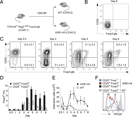
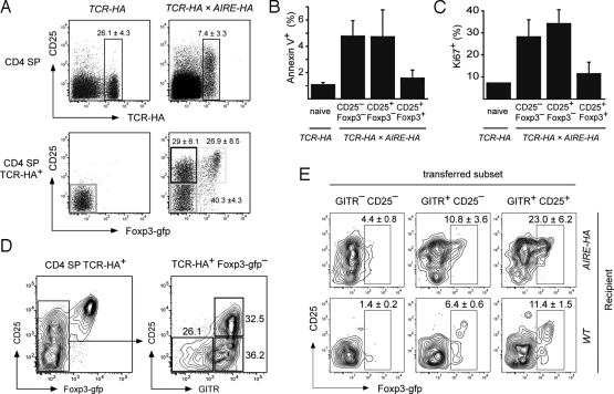
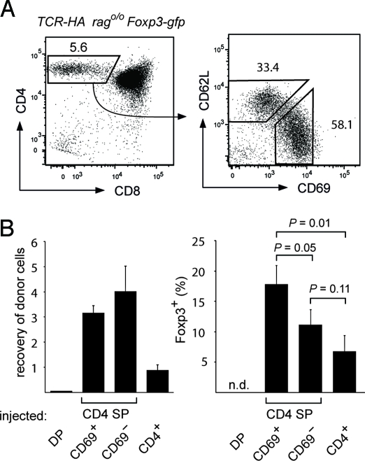
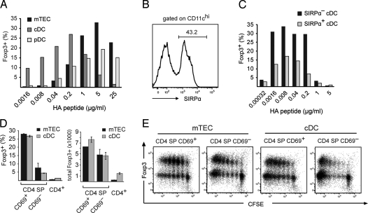
The majority of regulatory T cells (T(regs)) are believed to be of thymic origin. It has been hypothesized that this may result from unique intrathymic environmental cues, possibly requiring a dedicated antigen-presenting cell (APC). However, T cell-intrinsic developmental regulation of the susceptibility to T(reg) differentiation remains a mutually non-exclusive scenario. We found that upon exposure of monoclonal T cells of sequential developmental stages to a thymic microenvironment expressing cognate antigen, the efficiency of T(reg) induction inversely correlated with progressive maturation. This inclination of immature thymocytes toward T(reg) differentiation was even seen in an APC-free in vitro system, providing only TCR stimulation and IL-2. In support of quantitative but not qualitative features of external cues being critical, thymic epithelial cells as well as different thymic dendritic cell (DC)-subtypes efficiently induced T(reg) development of immature thymocytes, albeit at strikingly different optimal doses of cognate antigen. We propose that the intrinsically high predisposition of immature thymocytes to T(reg) development may contribute to the predominantly thymic origin of the T(reg) repertoire. The underlying instructive stimulus, however, does not require unique features of a dedicated APC and can be delivered by hematopoietic as well as epithelial thymic stromal cells.


Conflict of interest statement

The authors declare no conflict of interest.

Figures

Fig. 1.
Fig. 1.
Naïve TCR-HA+ cells give rise to Treg after intrathymic injection into AIRE-HA recipient mice. (A) Experimental design: 5 × 10⁵ CD4 SP cells from CD45.1 TCR-HA Rago/o Foxp3-gfp mice were intrathymically transferred into either WT or AIRE-HA recipients (CD45.2). (B) In WT recipients, transferred cells exhibited a stable Foxp3CD25 phenotype. (C) Kinetics of intrathymic Treg development in AIRE-HA thymi. Injected cells were analyzed for Foxp3-gfp and CD25 expression at different time points as indicated above the plots. Numbers in quadrants indicate the percentage (± SD, n = 3) of cells within the respective quadrant. (D) Emergence of CD25Foxp3+ and CD25+Foxp3+ cells. The diagram depicts the average percentage (± SD, n = 3) of CD25Foxp3+ or CD25+Foxp3+ recovered from AIRE-HA recipient mice at the indicated time points. (E) Recovery of injected cells. The diagram shows the average absolute number (± SD., n = 3) of cells recovered from intrathymically injected AIRE-HA or WT mice at the indicated time points. (F) Proliferation upon intrathymic antigen encounter. PKH26 labeled cells were i.t. injected into either WT or AIRE-HA recipient mice. The histogram shows an overlay of the 3 phenotypically distinct donor-derived subpopulations in AIRE-HA recipients and of total donor cells in WT recipient mice 5 days after i.t. injection. All data in Fig. 1 are representative of at least 3 independent experiments.
Fig. 2.
Fig. 2.
Phenotype and precursor/progeny relationship of HA-specific CD4 SP thymocytes in TCR-HA × AIRE-HA mice. (A) Staining of CD4 SP cells in 5-week-old TCR-HA or TCR-HA × AIRE-HA mice for TCR-HA and CD25 expression (Upper) or Foxp3-gfp and CD25 expression on gated TCR-HA+ CD4 SP cells (Lower). Numbers indicate the average frequency (± SD) of cells within gates. (n = 5 for TCR-HA mice, n = 35 for TCR-HA × AIRE-HA mice). (B and C) Foxp3-gfp negative TCR-HA+ CD4 SP cells of TCR-HA × AIRE-HA mice show increased rates of apoptosis and proliferation. The percentage of apoptotic (B) or dividing cells (C) as assessed by staining for Ki67 or Annexin V, respectively, is shown. (D and E) Foxp3 CD4 SP subpopulations of TCR-HA × AIRE-HA mice contain Treg precursors. Foxp3 cells of TCR-HA × AIRE-HA CD45.1 mice were sorted into the indicated subpopulations based upon CD25 and GITR expression (D) and i.t. transferred into AIRE-HA or WT mice. After 4 days, mice were killed and donor-derived thymocytes were analyzed for Foxp3-gfp and CD25 expression (E). Representative plots for each subset injected into AIRE-HA or WT mice are shown. Numbers in plots indicate the average frequency (± SD) of donor derived Foxp3-gfp+ cells recovered. Data are representative of 4 independent experiments.
Fig. 3.
Fig. 3.
The maturation stage of CD4 SP cells determines the efficiency of Treg conversion in vivo. (A) Gated TCR-HA Rag2o/o CD4 SP thymocytes (Left) can be subdivided into immature (CD69+CD62L) and mature (CD69CD62L+) cells (Right). Numbers indicate the percentage of cells in the respective gates. (B) Intrathymic transfer of thymocyte subpopulations and peripheral T cells. DP, CD69+CD62L CD4 SP, CD69CD62L+ CD4 SP, and peripheral CD4+ cells from TCR-HA Rago/o Foxp3-gfp mice (CD45.1) were sorted and mixed with a PKH26 labeled reference population of total CD4 SP cells (CD45.1) at a ratio of 1:2.6 before intrathymic transfer into AIRE-HA recipients. After 5 days, the recovery of the different tester populations was determined as the ratio of the respective cells to reference cells among donor cells (CD45.1+) (Left). The Right diagram shows the percentage of Foxp3-gfp+ cells among tester cells (n.d. = not detectable). Data are representative of 2 independent experiments with n = 3.
Fig. 4.
Fig. 4.
Induction of Treg cells by different thymic stromal APCs in vitro. (A) TCR-HA Rag2o/o CD4 SP thymocytes were co-cultured with mTECs, thymic cDCs, and thymic pDCs in the presence of increasing amounts of HA peptide. After 5 days, T cells were analyzed for CD25 and Foxp3-gfp expression. The bar diagram shows the percentage of Foxp3+ cells recovered. (B) Separation of thymic cDC into Sirpα+ and Sirpα subpopulations. The histogram shows a Sirpα staining of gated CD11chigh thymic dendritic cells (C) Sirpα+ and Sirpα DC were co-cultured with TCR-HA+ CD4 SP cells in the presence of increasing amounts of HA peptide. The diagram shows the percentage of Foxp3+ cells recovered after 5 days. (D) T cell maturation impinges on the efficiency of Treg induction in vitro. The indicated T cell subpopulations were co-cultured with mTEC or cDC at their respective optimal peptide concentrations. The diagrams show the percentage (Left) or total number (Right) of Foxp3+ cells recovered after 5 days. (E) Inverse correlation of Treg induction and proliferation. CFSE-labeled mature and immature CD4 SP cells from TCR-HA Rag2o/o mice were co-cultured with mTEC or cDC at their respective optimal peptide concentration and analyzed for Foxp3 expression and CFSE dilution after 5 days.
Fig. 5.
Fig. 5.
Treg induction in an APC-free system. (A) Immature and mature CD4 SP T cells from TCR-HA rago/o Foxp3-gfp mice were sorted and cultured in the presence of plate-bound anti-CD3. Cells were analyzed for CD25 and Foxp3-gfp expression after 3 days. The percentage (Left) and absolute numbers (Right) of Foxp3+ cells is depicted. (B) Immature (CD69+CD62L) and mature (CD69CD62L+) polyclonal CD25Foxp3 CD4 SP cells were sorted from Foxp3-gfp mice and cultured and analyzed as in A.

References

    1. Sakaguchi S. Naturally arising CD4+ regulatory T cells for immunologic self-tolerance and negative control of immune responses. Annu Rev Immunol. 2004;22:531–562. - PubMed
    1. Hsieh CS, Zheng Y, Liang Y, Fontenot JD, Rudensky AY. An intersection between the self-reactive regulatory and nonregulatory T cell receptor repertoires. Nat Immunol. 2006;7:401–410. - PubMed
    1. Pacholczyk R, Ignatowicz H, Kraj P, Ignatowicz L. Origin and T cell receptor diversity of Foxp3+CD4+CD25+ T cells. Immunity. 2006;25:249–259. - PubMed
    1. Wong J, Mathis D, Benoist C. TCR-based lineage tracing: No evidence for conversion of conventional into regulatory T cells in response to a natural self-antigen in pancreatic islets. J Exp Med. 2007;204:2039–2045. - PMC - PubMed
    1. Apostolou I, Sarukhan A, Klein L, von Boehmer H. Origin of regulatory T cells with known specificity for antigen. Nat Immunol. 2002;3:756–763. - PubMed

Publication types