. 2009 Jul 31;284(31):20796-803.
doi: 10.1074/jbc.M109.025353. Epub 2009 Jun 10.

Calcium elevation in mitochondria is the main Ca2+ requirement for mitochondrial permeability transition pore (mPTP) opening

Affiliations

Calcium elevation in mitochondria is the main Ca2+ requirement for mitochondrial permeability transition pore (mPTP) opening

Heidi K Baumgartner et al. J Biol Chem. .

Abstract

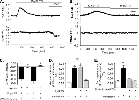
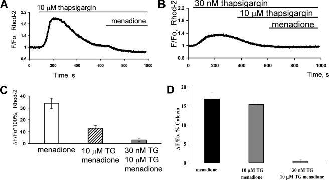
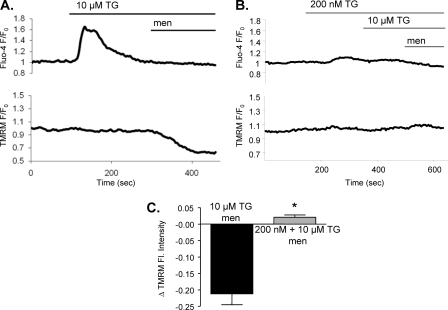
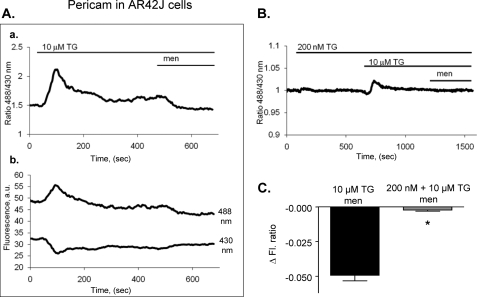
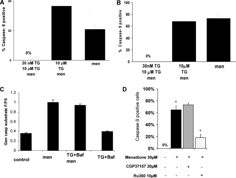
We have investigated in detail the role of intra-organelle Ca2+ content during induction of apoptosis by the oxidant menadione while changing and monitoring the Ca2+ load of endoplasmic reticulum (ER), mitochondria, and acidic organelles. Menadione causes production of reactive oxygen species, induction of oxidative stress, and subsequently apoptosis. In both pancreatic acinar and pancreatic tumor AR42J cells, menadione was found to induce repetitive cytosolic Ca2+ responses because of the release of Ca2+ from both ER and acidic stores. Ca2+ responses to menadione were accompanied by elevation of Ca2+ in mitochondria, mitochondrial depolarization, and mitochondrial permeability transition pore (mPTP) opening. Emptying of both the ER and acidic Ca2+ stores did not necessarily prevent menadione-induced apoptosis. High mitochondrial Ca2+ at the time of menadione application was the major factor determining cell fate. However, if mitochondria were prevented from loading with Ca2+ with 10 mum RU360, then caspase-9 activation did not occur irrespective of the content of other Ca2+ stores. These results were confirmed by ratiometric measurements of intramitochondrial Ca2+ with pericam. We conclude that elevated Ca2+ in mitochondria is the crucial factor in determining whether cells undergo oxidative stress-induced apoptosis.


Figures

FIGURE 1.
FIGURE 1.
A sharp increase in cytosolic calcium is required for induction of apoptosis in acinar cells. Isolated mouse pancreatic acinar cells were loaded with 100 nm TMRM-AM and 2.5 μm Fluo-4-AM (A and B). Fluorescence was measured over time before and after treatment with 30 μm menadione in the presence of 10 μm thapsigargin (A) or 30 nm + 10 μm thapsigargin (B). Changes of TMRM fluorescence after menadione treatment in different calcium store depletion conditions were compared (C). Separate groups of isolated cells were incubated with general caspase substrate, and cells were treated with 10 μm thapsigargin (D) or 30 nm and subsequent 10 μm thapsigargin (E) before application of 30 μm menadione (mean ± S.E., *, p < 0.05).
FIGURE 2.
FIGURE 2.
Menadione can induce release of calcium from mitochondria of acinar cells due to mPTP. Isolated mouse pancreatic acinar cells were loaded with Rhod 2-AM (A–C) or Calcein AM (D). Fluorescence was measured over time before and after treatment with menadione in the presence of 10 μm thapsigargin (A) or 30 nm thapsigargin followed by 10 μm thapsigargin (B). A comparison of amplitudes of changes of Rhod 2 fluorescence after menadione treatment under A and B conditions is shown in C (mean ± S.E., *, p < 0.05). D, fluorescence of calcein was quenched with CoCl2 in all intracellular organelles except mitochondria. Menadione induces mPTP as shown previously (8). Pretreatment for 10 min with high dose (10 μm) thapsigargin did not change the mPTP opening in response to menadione. However, pretreatment with low dose (30 nm) and subsequent high (10 μm) thapsigargin prevented mPTP induction by menadione (mean ± S.E., *, p < 0.05).
FIGURE 3.
FIGURE 3.
AR42J cells also require sharp increases in cytosolic calcium for induction of apoptosis. AR42J cells were loaded with 25 nm TMRM-AM and 10 μm Fluo-4-AM (A and B). Fluorescence was measured over time before and after application of 30 μm menadione in cells pretreated with 10 μm thapsigargin (A, n = 6) or 200 nm thapsigargin followed by 10 μm thapsigargin (B, n = 6). Changes in TMRM fluorescence after menadione treatment were compared in C (mean ± S.E., *, p < 0.05).
FIGURE 4.
FIGURE 4.
Menadione can induce release of calcium from mitochondria of AR42J cells (pericam measurements). AR42J cells were transfected with fluorescent mitochondrial ratiometric calcium pericam. Fluorescence was measured over time before and after menadione in cells pretreated for 10 min with 10 μm thapsigargin (A) or for 10 min with 200 nm and subsequently with 10 μm thapsigargin (B). The ratio of pericam fluorescence is shown in Aa and B, while original traces of pericam fluorescence (488 nm and 430 nm excitation) are shown in Ab. The change in fluorescence after menadione treatment under each condition (A and B) was compared (C) (mean ± S.E., n = 8–11 per group, *, p < 0.05).
FIGURE 5.
FIGURE 5.
Caspase-8 and -9 activation by menadione with different ways of emptying calcium stores in pancreatic acinar cells is shown. Caspase-8 (A) and -9 (B) activation by menadione when calcium content of ER was reduced slowly (30 nm thapsigargin followed by 10 μm thapsigargin) or quickly (10 μm thapsigargin) or with 100 nm Bafilomycin A1. Bars represent percent of apoptotic cells. C shows general caspase substrate controls in the presence of thapsigargin and Bafilomycin A1. D shows that caspase-9 activation is significantly inhibited by the mitochondrial uniporter inhibitor Ru360.

References

    1. Thornberry N. A., Lazebnik Y. (1998) Science 281, 1312–1316 - PubMed
    1. Berridge M. J., Lipp P., Bootman M. D. (2000) Nat. Rev. Mol. Cell Biol. 1, 11–21 - PubMed
    1. Huang Y., Fischer J. E., Balasubramaniam A. (1996) Peptides 17, 497–502 - PubMed
    1. Gerasimenko J. V., Gerasimenko O. V., Palejwala A., Tepikin A. V., Petersen O. H., Watson A. J. M. (2002) J. Cell Science 115, 485–497 - PubMed
    1. Baumgartner H. K., Gerasimenko J. V., Thorne C., Ashurst L. H., Barrow S. L., Chvanov M. A., Gillies S., Criddle D. N., Tepikin A. V., Petersen O. H., Sutton R., Watson A. J., Gerasimenko O. V. (2007) Am. J. Physiol. Gastrointest. Liver Physiol. 293, G296–G307 - PubMed

Publication types