. 2009 Jun 10;29(23):7519-25.
doi: 10.1523/JNEUROSCI.0517-09.2009.

Shedding light on serpent sight: the visual pigments of henophidian snakes

Affiliations

Shedding light on serpent sight: the visual pigments of henophidian snakes

Wayne L Davies et al. J Neurosci. .

Abstract

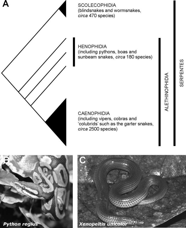
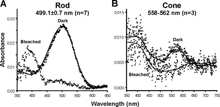
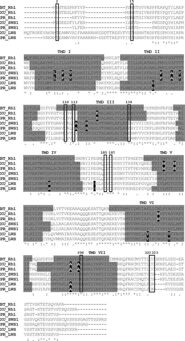
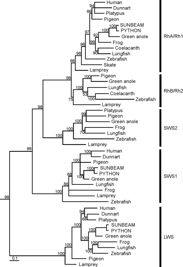
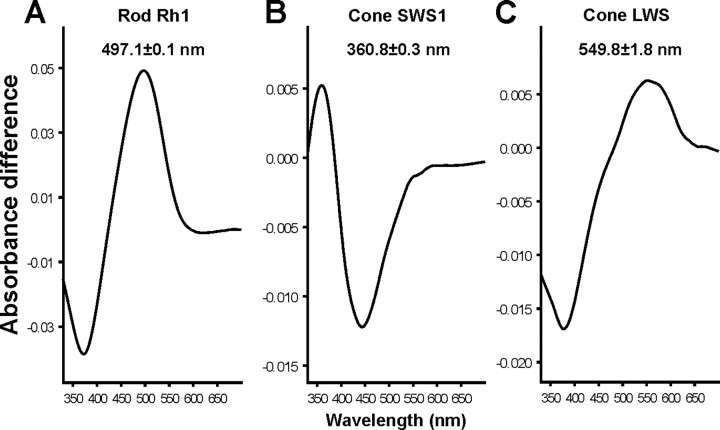
The biologist Gordon Walls proposed his "transmutation" theory through the 1930s and the 1940s to explain cone-like morphology of rods (and vice versa) in the duplex retinas of modern-day reptiles, with snakes regarded as the epitome of his hypothesis. Despite Walls' interest, the visual system of reptiles, and in particular snakes, has been widely neglected in favor of studies of fishes and mammals. By analyzing the visual pigments of two henophidian snakes, Xenopeltis unicolor and Python regius, we show that both species express two cone opsins, an ultraviolet-sensitive short-wavelength-sensitive 1 (SWS1) (lambda(max) = 361 nm) pigment and a long-wavelength-sensitive (LWS) (lambda(max) = 550 nm) pigment, providing the potential for dichromatic color vision. They also possess rod photoreceptors which express the usual rod opsin (Rh1) pigment with a lambda(max) at 497 nm. This is the first molecular study of the visual pigments expressed in the photoreceptors of any snake species. The presence of a duplex retina and the characterization of LWS, SWS1, and Rh1 visual pigments in henophidian snakes implies that "lower" snakes do not provide support for Walls' transmutation theory, unlike some "higher" (caenophidian) snakes and other reptiles, such as geckos. More data from other snake lineages will be required to test this hypothesis further.


Figures

Figure 1.
Figure 1.
A, A schematic cladogram showing the relationships between blindsnakes and wormsnakes (Scolecophidia), lower snakes (Henophidia) (e.g., pythons, boas, and sunbeam snakes), and higher snakes (Caenophidia) (e.g., vipers, cobras, and colubrids). B, C, Photograph of the head and upper body of two henophidian snakes, the royal python (Python regius) (B) and the Old World sunbeam snake (Xenopeltis unicolor) (C). Photographs courtesy of Patrick Jean, Muséum d'histoire naturelle de Nantes, France (P. regius) and Jodi Rowley, James Cook University, Australia (X. unicolor).
Figure 2.
Figure 2.
Mean absorbance spectra from microspectrophotometry of rods (A) and cones (B) of X. unicolor under dark and light-bleached conditions. The prebleach rod spectrum (open squares) is overlaid with a vitamin-A1 (rhodopsin) visual pigment template (line); cone spectra are fitted with variable point unweighted running averages.
Figure 3.
Figure 3.
Alignment of the amino acid sequences of visual pigments expressed in the retina of the sunbeam (X. unicolor) and python (P. regius) snakes. Amino acid numbering is based on the Rh1 pigment of the common cow (Bos taurus, BT). Asterisks denote an identical consensus residue between all snake opsin sequences and bovine Rh1. “:” and “.” indicate a conservative or semiconservative amino substitution, respectively, with the codon-matched protein alignment. Gaps inserted to maintain a high degree of identity present between the opsin sequences derived from both sunbeam and python snake retina and bovine Rh1 are indicated by dashes (−). Seven putative transmembrane domains (TMDs) are indicated by gray shading. The TMDs shown for bovine rhodopsin were determined by crystallography (Palczewski et al., 2000). The putative positions of the TMDs for each snake visual pigment were determined online using TMHMM Server Version 2.0 (http://www.cbs.dtu.dk/services/TMHMM/). Residues identified as being critical for correct opsin protein conformation are boxed. Comparison of the opsin amino acid sequences of X unicolor, P. regius, and bovine rod opsin demonstrated that the critical residues involved in the maintenance of the tertiary structure of the opsin molecule are present. Using the conventional numbering system of the bovine rod opsin polypeptide sequence, these key sites include (1) three conserved cysteine (C) residues at positions 110 (TMD3), 185 [extracellular domain (ECD2)], and 187 (ECD2) that are involved in disulfide bond formation (Karnik and Khorana, 1990), except for a Thr (T) residue at position 185 in the LWS opsin of both snakes, which is also conserved throughout the rest of the vertebrate LWS opsin class; (2) a conserved glutamate (E) at position 113 (TMD3) that provides the negative counterion to the proton of the Schiff base (Sakmar et al., 1989); (3) a conserved glutamate (E) at position 134 (TM3) that provides a negative charge to stabilize the inactive opsin molecule (Cohen et al., 1992); (4) a conserved lysine (K) at position 296 (TM7) that is covalently linked to the chromophore via a Schiff base (Dratz and Hargrave, 1983); (5) conservation of two cysteine (C) residues at putative palmitoylation positions 322 and 323 (Ovchinnikov et al., 1988) in both sunbeam and python snake Rh1 opsins but not LWS and SWS1 opsins; (6) the presence of a number of Ser (S) and Thr (T) residues in the carboxy terminus, which are potential targets for phosphorylation by rhodopsin kinases in the deactivation of metarhodopsin II (Palczewski et al., 1993; Zhao et al., 1997); and (7) the conserved glycosylation sites at positions 2 and 15 (Kaushal et al., 1994) in the Rh1 opsin identified in the retina of X. unicolor and P. regius. Amino acids important for the spectral tuning of LWS, SWS1, and Rh1 visual pigments are indicated as black highlighted boxes with white text.
Figure 4.
Figure 4.
Phylogenetic analysis of P. regius and X. unicolor LWS, SWS1, and Rh1 retinal opsins (GenBank accession numbers FJ497233-FJ497238) with LWS, SWS1, SWS2, RhB/Rh2, and RhA/Rh1 visual pigment coding sequences of other vertebrate species. Posterior probability values (represented as a percentage) are indicated for each resolved node. The scale bar indicates the number of nucleotide substitutions per site. The ciliary opsin of Dumeril's clam worm, Platynereis dumerilii, was used as an outgroup (data not shown). The sequences used for generating the tree are as follows: (1) RhA/Rh1 opsin class: human (Homo sapiens), NM000539; fat-tailed dunnart (Sminthopsis crassicaudata), AY159786; platypus (Ornithorhynchus anatinus), EF050076; pigeon (Columba livia), AH007730; sunbeam snake (Xenopeltis unicolor), FJ497233; python (Python regius), FJ497236; green anole (Anolis carolinensis), AOIRHODOPS; clawed frog (Xenopus laevis), NM001087048; Comoran coelacanth (Latimeria chalumnae), AF131253; Australian lungfish (Neoceratodus forsteri), EF526295; zebrafish (Danio rerio), NM131084; little skate (Raja erinacea), U81514; pouched lamprey (Geotria australis) (RhA), AY366493; (2) RhB/Rh2 opsin class: pigeon (Columba livia), AH007731; green anole (Anolis carolinensis), AH004781; Australian lungfish (Neoceratodus forsteri), EF526296; Comoran coelacanth (Latimeria chalumnae), AF131258; zebrafish (Danio rerio), NM131253 (Rh2.1); pouched lamprey (Geotria australis) (RhB), AY366494; (3) SWS2 opsin class; platypus (Ornithorhynchus anatinus), EF050077; pigeon (Columba livia), AH007799; green anole (Anolis carolinensis), AF133907; clawed frog (Xenopus laevis), BC080123; Australian lungfish (Neoceratodus forsteri), EF526299; zebrafish (Danio rerio), NM131192; pouched lamprey (Geotria australis), AY366492; (4) SWS1 opsin class: human (Homo sapiens), NM001708; fat-tailed dunnart (Sminthopsis crassicaudata), AY442173; pigeon (Columba livia), AH007798; sunbeam snake (Xenopeltis unicolor), FJ497234; python (Python regius), FJ497237; green anole (Anolis carolinensis), AH007736; Australian lungfish (Neoceratodus forsteri), EF526298; clawed frog (Xenopus laevis), XLU23463; pouched lamprey (Geotria australis), AY366495; zebrafish (Danio rerio), NM131319; (5) LWS opsin class: human (Homo sapiens), NM020061; fat-tailed dunnart (Sminthopsis crassicaudata), AY430816; platypus (Ornithorhynchus anatinus), EF050078; sunbeam snake (Xenopeltis unicolor), FJ497235; python (Python regius), FJ497238; green anole (Anolis carolinensis), ACU08131; clawed frog (Xenopus laevis), XLU90895; Australian lungfish (Neoceratodus forsteri), EF526297; zebrafish (Danio rerio), NM131175; pigeon (Columba livia), AH007800; pouched lamprey (Geotria australis), AY366491.
Figure 5.
Figure 5.
Difference absorbance spectra for the Rh1 (A), SWS1 (B), and LWS (C) visual pigments of X. unicolor, regenerated in vitro with 11-cis-retinal.

Similar articles

Cited by

References

    1. Bowmaker JK, Astell S, Hunt DM, Mollon JD. Photosensitive and photostable pigments in the retinae of Old World monkeys. J Exp Biol. 1991;156:1–19. - PubMed
    1. Buck SL. Influence of rod signals on hue perception: evidence from successive scotopic contrast. Vision Res. 1997;37:1295–1301. - PubMed
    1. Cohen GB, Oprian DD, Robinson PR. Mechanism of activation and inactivation of opsin: role of Glu113 and Lys296. Biochemistry. 1992;31:12592–12601. - PubMed
    1. Conant R, Collins J. Boston: Houghton Mifflin; 1998. A field guide to reptiles and amphibians of eastern and central North America (Peterson field guide series)
    1. Cowing JA, Poopalasundaram S, Wilkie SE, Robinson PR, Bowmaker JK, Hunt DM. The molecular mechanism for the spectral shifts between vertebrate ultraviolet- and violet-sensitive cone visual pigments. Biochem J. 2002;367:129–135. - PMC - PubMed

Publication types

MeSH terms

LinkOut - more resources