. 2009 Jun 10;29(23):7549-57.
doi: 10.1523/JNEUROSCI.2474-08.2009.

Exercise induces cortical plasticity after neonatal spinal cord injury in the rat

Affiliations

Exercise induces cortical plasticity after neonatal spinal cord injury in the rat

Tina Kao et al. J Neurosci. .

Abstract

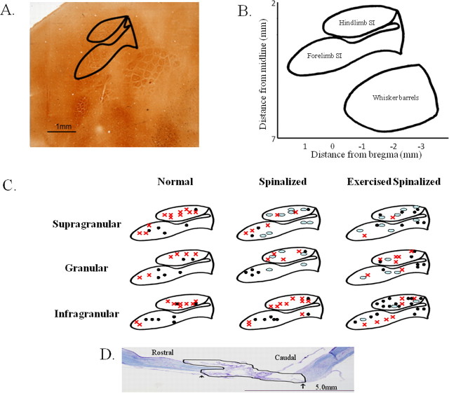
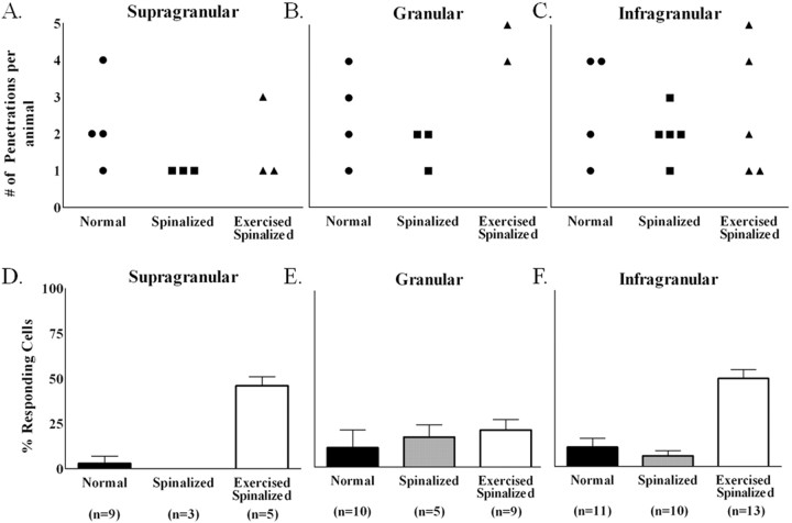
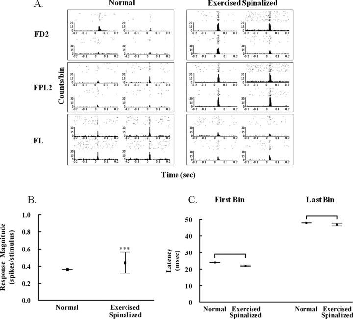
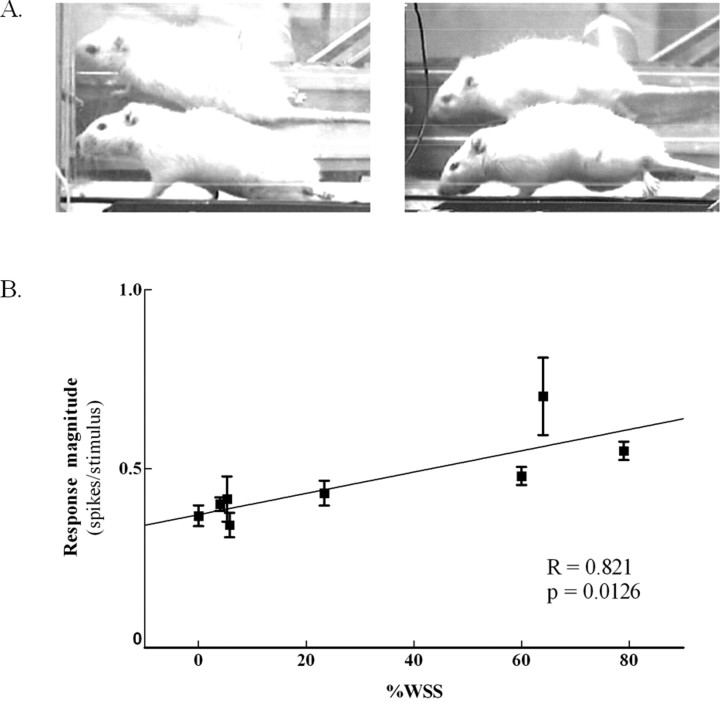
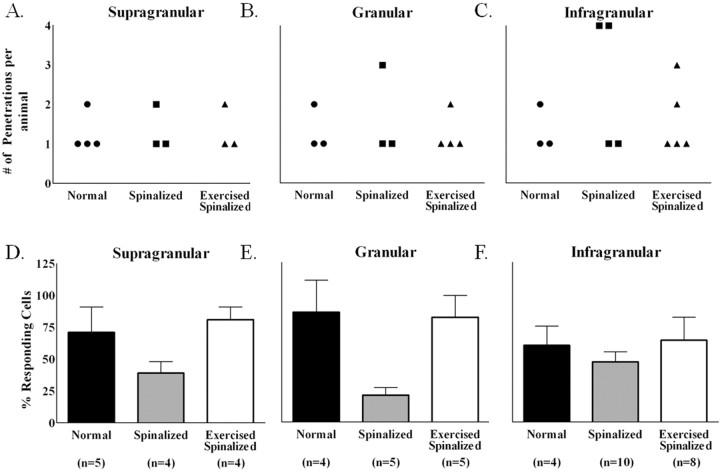
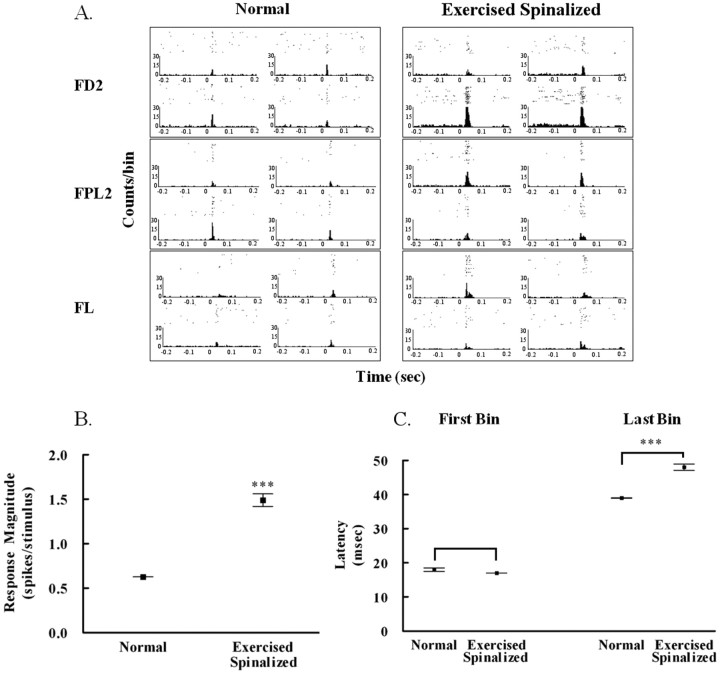
Exercise-induced cortical plasticity is associated with improved functional outcome after brain or nerve injury. Exercise also improves functional outcomes after spinal cord injury, but its effects on cortical plasticity are not known. The goal of this investigation was to study the effect of moderate exercise (treadmill locomotion, 3 min/d, 5 d/week) on the somatotopic organization of forelimb and hindlimb somatosensory cortex (SI) after neonatal thoracic transection. We used adult rats spinalized as neonates because some of these animals develop weight-supported stepping, and, therefore, the relationship between cortical plasticity and stepping could also be examined. Acute, single-neuron mapping was used to determine the percentage of cortical cells responding to cutaneous forelimb stimulation in normal, spinalized, and exercised spinalized rats. Multiple single-neuron recording from arrays of chronically implanted microwires examined the magnitude of response of these cells in normal and exercised spinalized rats. Our results show that exercise not only increased the percentage of responding cells in the hindlimb SI but also increased the magnitude of the response of these cells. This increase in response magnitude was correlated with behavioral outcome measures. In the forelimb SI, neonatal transection reduced the percentage of responding cells to forelimb stimulation, but exercise reversed this loss. This restoration in the percentage of responding cells after exercise was accompanied by an increase in their response magnitude. Therefore, the increase in responsiveness of hindlimb SI to forelimb stimulation after neonatal transection and exercise may be due, in part, to the effect of exercise on the forelimb SI.


Figures

Figure 1.
Figure 1.
Penetration sites for the acute single-neuron mapping of forelimb and hindlimb somatosensory cortex in normal, spinalized, and exercised spinalized rats. A, The flattened somatosensory cortex, when stained with cytochrome oxidase in a normal adult rat, reveals the locations that we targeted in this study. Solid lines outline the hindlimb and forelimb somatosensory cortices. B, Schematic of the flattened somatosensory cortex based on 1 mm grids showing the stereotaxic coordinates of the forelimb and hindlimb somatosensory cortex. C, Penetration sites into the forelimb and hindlimb somatosensory cortex for each group of rats. Responses were recorded from electrodes placed in the supragranular, granular, and infragranular layers of the hindlimb and forelimb somatosensory cortices. Black circles, Electrode penetration sites that elicited cells which responded to sensory stimulation. Red x, Electrode penetration sites that elicited cells which did not respond to sensory stimulation. Ovals, Electrode penetration sites that did not elicit any cells. D, Nissl/myelin staining of the spinal cord of a representative adult rat transected as a neonate at the T8/T9 level. No cell bodies or axons were observed in the transection site (outlined in black). The extent of this representative injury was comparable with the other injuries. The average rostral to caudal extent of the injury was 6.4 ± 2.2 mm, and no differences were observed in the extent of the injury between the spinalized and exercised spinalized groups. Scale bar, 5.0 mm.
Figure 2.
Figure 2.
Exercise increased the percentages of cells in the extragranular layers of the hindlimb somatosensory cortex of spinalized rats that responded to touch stimulation of the forelimb periphery. Data were collected from four normal, five spinalized, and five exercised spinalized rats. A–C, The numbers of electrode penetrations within the hindlimb somatosensory cortex for which cells were detected are shown for each animal (circles represent normal animals, squares represent spinalized animals, and triangles represent exercised spinalized animals). D–F, The averages of the percentage of cells that responded to sensory stimuli for each penetration (n = total number of penetrations) are plotted as a per layer basis for each animal group. As expected, a small number of cells within the hindlimb somatosensory cortex responded to touch stimulation of the forelimb periphery in all layers of normal rats: [supragranular (3% of 64 cells), granular (11% of 55 cells), and infragranular (11% of 192 cells)]. After neonatal T8/T9 transection, the percentages of cells that responded to touch stimulation did not change when compared with those of normal rats [supragranular (0% of 15 cells), granular (17% of 24 cells), and infragranular (6% of 111 cells)]. Exercise increased the percentages of sensory responsive cells in both the supragranular (46% of 44 cells) and infragranular (50% of 211 cells) layers of spinalized rats when compared with those of the normal and nonexercised spinalized rats. Exercise did not increase the percentage of cells responding to touch stimulation in the granular layer (21% of 39 cells) of spinalized rats when compared with those of the normal and the nonexercised spinalized rats. Error bars indicate SEM.
Figure 3.
Figure 3.
Exercise increased the response magnitude of cells in the infragranular hindlimb cortex to forelimb stimulation in spinalized rats. A, Examples of peristimulus time histograms showing the responses of four infragranular hindlimb cells recorded simultaneously from each of three stimulus locations: forepaw digit 2 (FD2), forepaw palm pad 5 (FPL5), and forelimb (FL). B, The average response magnitude of infragranular cells within the hindlimb somatosensory cortex of exercised spinalized rats (0.4408 ± 0.12 spikes/stimulus, N = 10 rats) was greater than that of normal rats (0.3625 ± 0.002 spikes/stimulus, N = 9 rats); ***p < 0.001. C, No differences were observed between the first bin latency of exercised spinalized rats (22 ± 0.4 ms) versus that of normals (24 ± 0.08 ms) or the last bin latency of exercised spinalized rats (47 ± 0.8 ms) versus that of normals (48 ± 0.13 ms). Error bars indicate SEM.
Figure 4.
Figure 4.
The response magnitude of cells in the infragranular layer of the hindlimb cortex correlated with percentage weight-supported steps in exercised spinalized rats. A, Photographs of two adult rats with neonatal T8/T9 transections performing treadmill exercise (a mirror is placed behind the treadmill to ensure all activity of the hindlimbs is captured). On the left is one exercised spinalized rat exhibiting no weight support. The hindquarters are dragged on the surface of the treadmill. On the right is an exercised spinalized rat exhibiting a weight-supported step. The hindlimbs are supporting the hindquarters above the surface of the treadmill. B, The average weight-supported steps for eight rats were obtained after electrode implantation in the hindlimb somatosensory cortex. Each box represents different days of the sensory maps. Five rats had undergone two different days of sensory maps, whereas three rats had one sensory map. The average response magnitude of the cells were positively correlated with %WSS (**p < 0.01).
Figure 5.
Figure 5.
Exercise restored the percentage of cells that responded to sensory stimulations in the forelimb somatosensory cortex of spinalized rats. Data were collected from four normal, four spinalized, and five exercised spinalized rats. A–C, The numbers of electrode penetrations within the forelimb somatosensory cortex from which cells were detected are shown for each animal (circles represent normal animals, squares represent spinalized animals, and triangles represent exercised spinalized animals). D–F, The averages of the percentages of cells that responded to sensory stimuli for each penetration (n = total number of penetrations) were plotted as a per layer basis for each animal group. As expected, a high percentage of cells in all cortical layers of the forelimb cortex responded to sensory stimulation of the forelimb [supragranular (71% of 42 cells), granular (86% of 21 cells), and infragranular (60% of 77 cells)]. Neonatal spinalization decreased the percentages of sensory responsive cells in all layers of the forelimb cortex [supragranular (39% of 31 cells), granular (21% of 28 cells), and infragranular (47% of 181 cells)] when compared with those of the normal rats. Exercise restored the percentages of sensory responsive cells in all cortical layers of spinalized rats, comparable with those of normals [supragranular (81% of 21 cells), granular (82% of 27 cells), and infragranular (64% of 90 cells)]. Error bars indicate SEM.
Figure 6.
Figure 6.
Exercise increased the response magnitude of cells in the forelimb somatosensory cortex to forelimb stimulation in spinalized rats. A, Examples of peristimulus time histograms showing the responses of four cells recorded simultaneously from each of three stimulus locations: forepaw digit 2 (FD2), forepaw palm pad 5 (FPL5), and forelimb (FL). B, The average response magnitude of infragranular cells within the forelimb somatosensory cortex of exercised spinalized rats (1.49 ± 0.07 spikes/stimulus, N = 5 rats) was greater than that of normal rats (0.626 ± 0.001 spikes/stimulus, N = 4 rats); ***p < 0.001. C, There was no difference in the first bin latency of cortical forelimb cells of the exercised spinalized rats (17 ± 0.02 ms) when compared with the first bin latency from the normals (18 ± 0.47 ms). There was an increase in the last bin latency of the response of cortical cells in the exercised spinalized rats (48 ± 0.94 ms) when compared with that of the normals (39 ± 0.05 ms); ***p < 0.001. Error bars indicate SEM.

Similar articles

Cited by

References

    1. Abraham WC, Bear MF. Metaplasticity: the plasticity of synaptic plasticity. Trends Neurosci. 1996;19:126–130. - PubMed
    1. Cai LL, Fong AJ, Otoshi CK, Liang Y, Burdick JW, Roy RR, Edgerton VR. Implications of assist-as-needed robotic step training after a complete spinal cord injury on intrinsic strategies of motor learning. J Neurosci. 2006;26:10564–10568. - PMC - PubMed
    1. Chapin JK. Laminar differences in sizes, shapes, and response profiles of cutaneous receptive fields in the rat SI cortex. Exp Brain Res. 1986;62:549–559. - PubMed
    1. Chapin JK, Sadeq M, Guise JL. Corticocortical connections within the primary somatosensory cortex of the rat. J Comp Neurol. 1987;263:326–346. - PubMed
    1. Chau CW, McKinley PA. Chronological observations of primary somatosensory cortical maps in kittens following low thoracic (T12) spinal cord transection at 2 weeks of age. Somatosens Mot Res. 1991;8:355–376. - PubMed

Publication types