. 2009 Jun 10;29(23):7558-68.
doi: 10.1523/JNEUROSCI.0514-09.2009.

The unitary event underlying multiquantal EPSCs at a hair cell's ribbon synapse

Affiliations

The unitary event underlying multiquantal EPSCs at a hair cell's ribbon synapse

Geng-Lin Li et al. J Neurosci. .

Abstract

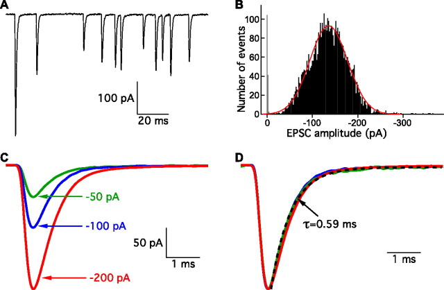
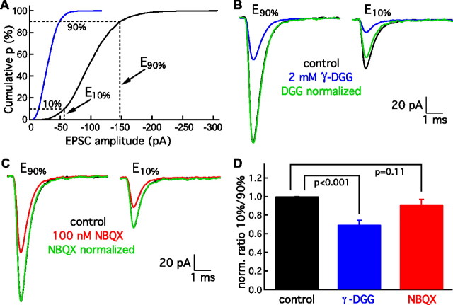
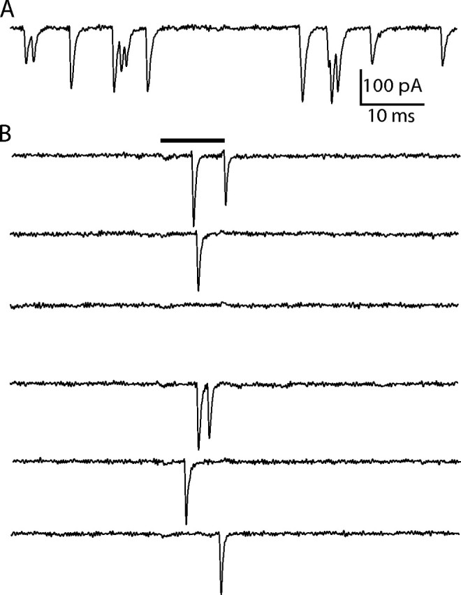
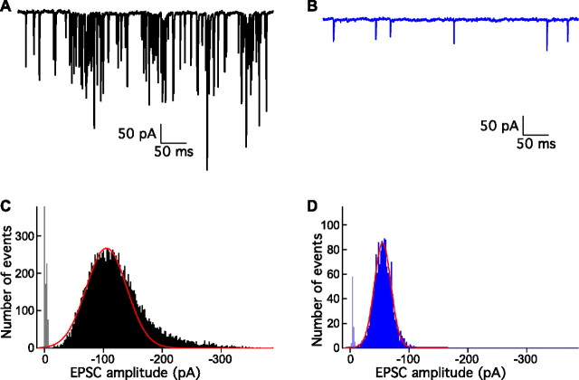
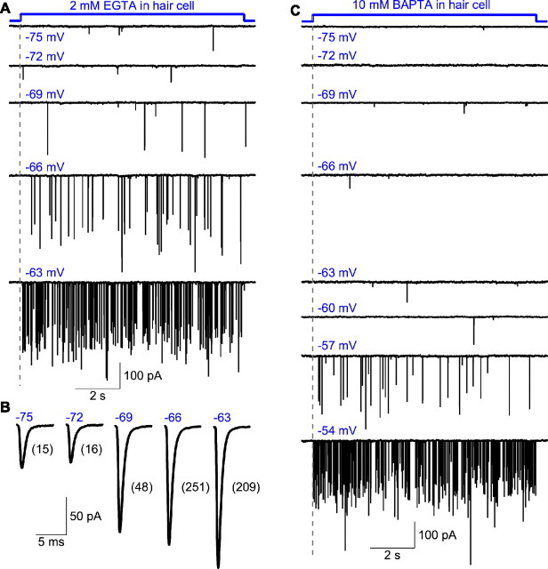
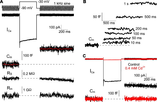
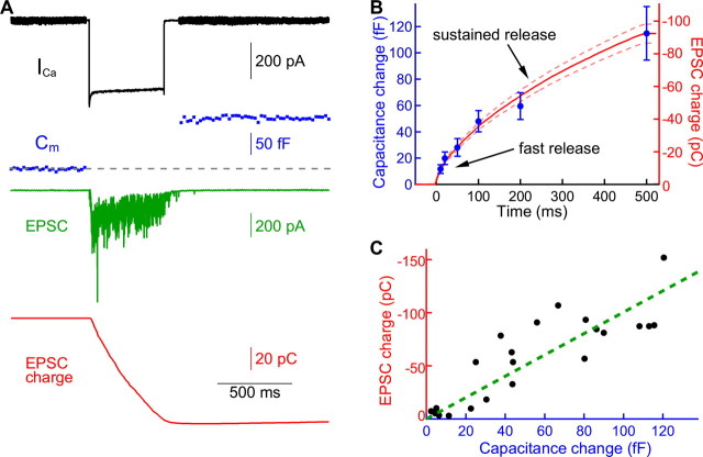
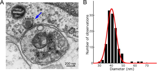
EPSCs at the synapses of sensory receptors and of some CNS neurons include large events thought to represent the synchronous release of the neurotransmitter contained in several synaptic vesicles by a process known as multiquantal release. However, determination of the unitary, quantal size underlying such putatively multiquantal events has proven difficult at hair cell synapses, hindering confirmation that large EPSCs are in fact multiquantal. Here, we address this issue by performing presynaptic membrane capacitance measurements together with paired recordings at the ribbon synapses of adult hair cells. These simultaneous presynaptic and postsynaptic assays of exocytosis, together with electron microscopic estimates of single vesicle capacitance, allow us to estimate a single vesicle EPSC charge of approximately -45 fC, a value in close agreement with the mean postsynaptic charge transfer of uniformly small EPSCs recorded during periods of presynaptic hyperpolarization. By thus establishing the magnitude of the fundamental quantal event at this peripheral sensory synapse, we provide evidence that the majority of spontaneous and evoked EPSCs are multiquantal. Furthermore, we show that the prevalence of uniquantal versus multiquantal events is Ca2+ dependent. Paired recordings also reveal a tight correlation between membrane capacitance increase and evoked EPSC charge, indicating that glutamate release during prolonged hair cell depolarization does not significantly saturate or desensitize postsynaptic AMPA receptors. We propose that the large EPSCs reflect the highly synchronized release of multiple vesicles at single presynaptic ribbon-type active zones through a compound or coordinated vesicle fusion mechanism.


Figures

Figure 1.
Figure 1.
Spontaneous EPSCs. A, Spontaneous EPSCs recorded from an afferent fiber exhibit a broad range of amplitudes. B, The amplitude distribution of 4884 spontaneous EPSCs is fit well with a Gaussian function (red). The average EPSC amplitude is −137 pA. The noise amplitude distribution is shown in gray. C, Averages of 10–20 EPSCs with amplitudes of −50 ± 2 pA (green), −100 ± 2 pA (blue), and −200 ± 2 pA (red) show that events of these different size classes possess smooth rise and decay phases. D, Normalization of the three groups of EPSCs in C reveals that they have similar kinetics. The dashed black line is a single-exponential fit to the decay phase of the −50 pA group. Traces in A–C are all from the same afferent fiber recording.
Figure 2.
Figure 2.
Differential block of EPSCs by γ-DGG. A, The cumulative amplitude distribution of EPSCs allows the selection of events in the smallest decile (E10%) and those in the largest decile (E90%). The blue trace shows that 2 mm γ-DGG, a low-affinity AMPA receptor antagonist, reduces the amplitudes of both small and large events. B, After averaging 10–20 events each from the E90% and E10% classes, events within ±2 pA, under control circumstances (black) or in the presence of 2 mm γ-DGG (blue), the result for the E90% class in γ-DGG is normalized to that in control solution (green). When the same normalization factor is applied to the E10% results, that for γ-DGG (green) is substantially smaller than that in control solution (black), indicating that γ-DGG blocks smaller EPSCs more efficaciously. C, In a comparison of EPSCs in control solution (black) with those in 100 nm NBQX (red), normalization shows that events in the E90% and E10% classes are blocked to a similar extent (green). D, When the ratios (E10%/E90%) for results in γ-DGG (blue) and NBQX (red) are normalized to their corresponding controls, the normalized ratio is significantly reduced by γ-DGG (6 cells) but not by NBQX (5 cells). Statistical significance was evaluated with a paired Student's t test.
Figure 3.
Figure 3.
Ca2+ iontophoresis onto a single active zone. A, Under control recording conditions, spontaneous EPSCs in an afferent fiber recording display a wide range of amplitudes. B, When the preparation is bathed in a zero Ca2+ solution, the spontaneous events are suppressed. A 10 ms local iontophoresis of Ca2+ (black bar) onto a putatively single synapse evokes EPSCs as large as those recorded from the same fiber in control solution. Failures to evoke EPSCs occurred for 37% of the iontophoretic applications at this site.
Figure 4.
Figure 4.
Presynaptic hyperpolarization during paired recordings. A, Spontaneous EPSC events originating from an unclamped hair cell display a large range of amplitudes. B, Voltage clamping the presynaptic hair cell to −90 mV eliminates all large events recorded from the same afferent fiber and reduces the frequency of the events. C, The rising phase of the amplitude distribution of 13,791 EPSCs from A was fit with a Gaussian function. Skewing of the distribution by large events occurred in a minority of recordings. The Gaussian fit yields a mean value of −105 pA, smaller than the value of −125 pA obtained from the average of all the events. D, Holding the hair cell at −90 mV shifted the amplitude distribution of 1542 EPSCs to a peak at −57 pA. The distribution is well fit by a Gaussian function with a mean value of −55 pA.
Figure 5.
Figure 5.
Ca2+ sensitivity of multiquantal EPSCs. A, EPSCs were recorded from a voltage-clamped afferent fiber, while a connected hair cell dialyzed with 2 mm EGTA was depolarized for 10 s from a holding potential of −90 mV to each of the indicated membrane potentials. The majority of the EPSCs recorded at the most negative potentials were small and putatively uniquantal, but depolarization progressively increased the proportion of large and presumably multiquantal events. B, Averaged EPSCs from the same fiber with the hair cell held at the membrane potentials indicated. The numbers in brackets indicate the number of EPSCs averaged at each hair cell membrane potential. The frequency and average amplitude of the EPSCs increased as the hair cell was depolarized more strongly. C, EPSCs were recorded from another afferent fiber, while a connected hair cell dialyzed with 10 mm BAPTA was stepped for 10 s from a holding potential of −90 mV to each of the indicated potentials. The presence of a fast Ca2+ buffer shifted the voltage dependence by ∼10 mV with respect to that in A. The records were obtained 3.5 min after the inception of the whole-cell recording from the hair cell.
Figure 6.
Figure 6.
Presynaptic capacitance measurements. A, A voltage-clamped hair cell was depolarized from a holding potential of −90 mV to −30 mV to evoke exocytosis. During the depolarizing stimulus, a presynaptic Ca2+ current (ICa) was recorded. A 1 kHz sinusoidal voltage 25 mV in amplitude was superposed on the holding potential before and after the depolarization. From the measured current response to this signal, the membrane capacitance (CM), series resistance (RS), and membrane resistance (RM) were calculated. The increase in capacitance reflects a rise in membrane surface area because of the fusion of synaptic vesicles. The red trace is the averaged value of the capacitance data points. Note that the series resistance and membrane resistance remain constant. B, The capacitance increases with the duration of the depolarizing stimulus. The traces shown are responses to single depolarizing pulses presented to the cell of panel A. C, Cd2+ at a concentration of 0.4 mm blocked both the Ca2+ current and the capacitance increase.
Figure 7.
Figure 7.
Paired recordings with capacitance measurements. A, In a representative hair cell afferent fiber paired recording, depolarization of the hair cell from −90 to −30 mV elicited a presynaptic Ca2+ current (black) and capacitance increase (blue) and a postsynaptic EPSC (green). The total charge transferred (red) is the integral of the evoked EPSC. B, The postsynaptic charge transferred (red, n = 11; dashed lines indicate SE range) is well correlated with the increase in capacitance of the presynaptic membrane (blue; n = 6) induced by depolarizing pulses of different durations. This agreement suggests that AMPA receptors are not significantly saturated or desensitized during exocytosis. C, For each paired recording, the total postsynaptic charge transferred was plotted against the presynaptic membrane capacitance increase. The data were fit with a straight line through the origin with a slope of 1.01 kC · F−1 and a correlation coefficient of 0.86.
Figure 8.
Figure 8.
Electron microscopic analysis of synaptic vesicle diameters. A, An electron microscopic image of a hair cell synapse shows a presynaptic active zone demarcated by its vesicle-endowed synaptic ribbon (blue arrow). The single postsynaptic afferent fiber branch shown in cross section contains mitochondria and several organelles. B, The amplitude distribution of synaptic vesicle diameters is fit well by a Gaussian function (red) with a mean of 40.3 nm.

References

    1. Albillos A, Dernick G, Horstmann H, Almers W, Alvarez de Toledo G, Lindau M. The exocytotic event in chromaffin cells revealed by patch amperometry. Nature. 1997;389:509–512. - PubMed
    1. Angleson JK, Betz WJ. Monitoring secretion in real time: capacitance, amperometry, fluorescence compared. Trends Neurosci. 1997;20:281–287. - PubMed
    1. Auger C, Marty A. Quantal currents at single-site central synapses. J Physiol. 2000;526:3–11. - PMC - PubMed
    1. Bekkers JM, Stevens CF. Cable properties of cultured hippocampal neurons determined from sucrose-evoked miniature EPSCs. J Neurophysiol. 1996;75:1250–1255. - PubMed
    1. Christie JM, Jahr CE. Multivesicular release at Schaffer collateral-CA1 hippocampal synapses. J Neurosci. 2006;26:210–216. - PMC - PubMed

Publication types

MeSH terms