. 2009 Jun 10;29(23):7582-90.
doi: 10.1523/JNEUROSCI.1336-09.2009.

Role of amyloid-beta glycine 33 in oligomerization, toxicity, and neuronal plasticity

Affiliations

Role of amyloid-beta glycine 33 in oligomerization, toxicity, and neuronal plasticity

Anja Harmeier et al. J Neurosci. .

Abstract

The aggregation of the amyloid-beta (Abeta) peptide plays a pivotal role in the pathogenesis of Alzheimer's disease, as soluble oligomers are intimately linked to neuronal toxicity and inhibition of hippocampal long-term potentiation (LTP). In the C-terminal region of Abeta there are three consecutive GxxxG dimerization motifs, which we could previously demonstrate to play a critical role in the generation of Abeta. Here, we show that glycine 33 (G33) of the central GxxxG interaction motif within the hydrophobic Abeta sequence is important for the aggregation dynamics of the peptide. Abeta peptides with alanine or isoleucine substitutions of G33 displayed an increased propensity to form higher oligomers, which we could attribute to conformational changes. Importantly, the oligomers of G33 variants were much less toxic than Abeta(42) wild type (WT), in vitro and in vivo. Also, whereas Abeta(42) WT is known to inhibit LTP, Abeta(42) G33 variants had lost the potential to inhibit LTP. Our findings reveal that conformational changes induced by G33 substitutions unlink toxicity and oligomerization of Abeta on the molecular level and suggest that G33 is the key amino acid in the toxic activity of Abeta. Thus, a specific toxic conformation of Abeta exists, which represents a promising target for therapeutic interventions.


Figures

Figure 1.
Figure 1.
Single amino acid exchanges within the GxxxG motif of Aβ42 modulate oligomerization. A, Amino acid sequence of Aβ1–42 with the glycines of the three consecutive GxxxG motifs numbered. The amino acid substitutions of the analyzed central GxxxG motif are in bold. B–F, SEC and Western blot analysis of freshly dissolved synthetic Aβ42 peptides. B, Aβ42 WT peptides preferentially form tetramers but also lower and higher oligomers. C, Aβ42 G29A peptides preferentially form tetramers and only few other oligomers. D, Aβ42 G33A variant peptide mainly form higher oligomers but also low-n oligomers. E, Aβ42 G33I substitution peptides almost exclusively form higher oligomers but hardly any monomers to decamers. F, Size separation of Aβ42 G29/33A yields high amounts of tetramers but also large amounts of high oligomers. Representative chromatograms of at least three independent measurements are shown. Western blots of freshly dissolved synthetic Aβ aggregates (Load) (compare figure insets) in the presence of SDS reveal monomers and high-n oligomers for all peptides. Compared with Aβ42 WT, G29A, and G29/33A, solely the amount of low-n oligomers of Aβ42 G33 substitution peptides is reduced.
Figure 2.
Figure 2.
Amino acid exchanges at position G29 and G33 affect accessibility of Aβ K28. A, MALDI-MS spectra of 3 h tryptic digests yield the following mass peaks (in Da for WT): Aβ1–42, 4512.3; Aβ1–28, 3261.4; Aβ17–42, 2576.3. Note that the mass shifts are because of amino acid substitutions. B, Generation of Aβ fragment 1–28 by limited tryptic digestion of Aβ42 WT, Aβ42 G29A, and Aβ42 G29/33A and (C) Aβ42 WT, Aβ42 G33A, and Aβ42 G33I. The amount of fragment 1–28 was calculated as the ratio (A1–28/A1–42) of ion peak areas of MALDI-MS spectra recorded at different time points. Note the different scale bars in B and C. Compared with Aβ42 WT, a rapid formation of high amounts of Aβ1–28 is observed when G29 is substituted but hardly any cleavage of trypsin at position K28 when G33 is exchanged. Representative spectra and evaluation of at least three independent measurements are shown.
Figure 3.
Figure 3.
Single amino acid exchange at position G33 modifies conformation of Aβ42 oligomers. Amino acid sequences and computational models of Aβ42 WT (A), Aβ42 G29A (B), Aβ42 G33A (C), Aβ42 G33I (D), and Aβ42 G29/33A (E) monomers and tetramers. Glycine residues 25, 29, 33, and 37 of the three consecutive GxxxG motifs are numbered, and substitutions at position G29 and G33 are in bold (sequence) and with arrows (monomer model). Negatively charged surfaces in red, positively charged in blue (Baker et al., 2001). Increase in hydrophobicity at G33 leads to stabilization of the folding core, provokes β-sheet conformation and increases oligomerization.
Figure 4.
Figure 4.
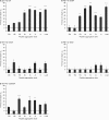
42 WT but not Aβ42 G33 substitution peptides are toxic to neuroblastoma cells. For all measurements, equal amounts of freshly dissolved peptides (Load) or of oligomer fractions after SEC and as determined by the BCA assays were used. Toxicity is described as percentage of cell death compared with untreated control cells (n = 4–8 ± SEM). A, Aβ42 WT exhibits a maximal toxicity. Toxicity increases from monomeric to tetrameric forms and then gradually decreases for higher oligomers. B, Aβ42 G29A reveals toxicity similar to the WT peptide. C, D, Both Aβ42 G33A (C) and Aβ42 G33I (D) lack any significant toxicity. E, Aβ42 G29/33A occupies an intermediate toxicity for the unseparated peptide. The toxicity of the Aβ42 G29/33A SEC fractions is similar to the WT for 4-mer, 10-mer, and 20-mer and in the range of Aβ42 G33I in case of the monomer, dimer, 6-mer, and 16-mer. A one-way ANOVA using Dunnett's multiple comparison test comparing peptides with untreated control was performed (*p < 0.01, **p < 0.001, ***p < 0.0001).
Figure 5.
Figure 5.
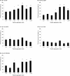
42 WT but not Aβ42 G33 substitution peptides are toxic to primary neurons. Measurements were performed with neurons incubated with equal amounts of freshly dissolved synthetic peptides (Load) or SEC fractions. Results are expressed as percentage of cell death. A, Aβ42 WT (n = 4–8 ± SEM) induces a maximal toxicity. Comparable with the data generated from neuroblastoma cells, toxicity increases from monomeric to tetrameric forms and then gradually decreases again as the peptide aggregates further into higher oligomers up to 20-mers. B, Similar to Aβ42 WT, the same pattern could be observed for Aβ42 G29A (n = 2–8 ± SEM). C, D, Aβ42 G33A (C) and Aβ42 G33I (D) (n = 4–8 ± SEM) cause no significant toxicity in primary neurons. E, Toxicity of Aβ42 G29/33A (n = 2–8 ± SEM) is similar to Aβ42 WT for monomers to tetramers and to Aβ42 G33 variant peptides for higher aggregates. A one-way ANOVA using Dunnett's multiple comparison test comparing peptides with untreated control was performed (*p < 0.001, **p < 0.0001).
Figure 6.
Figure 6.
42 WT but not Aβ42 G33 mutants are toxic in vivo in transgenic Drosophila melanogaster expressing extracellular Aβ42 WT or mutant peptides. For the analyses, strains of flies were selected for expressing equal amounts of Aβ forms (supplemental Fig. S3, available at www.jneurosci.org as supplemental material). Analysis of Aβ toxicity in transgenic flies by scanning electron microscopy of eye structure (top) and sections (bottom). A–D, Eyes of control flies (A) and flies expressing Aβ42 G33I (D) show a negligible distortion compared with those expressing Aβ42 WT (B) or Aβ42 G29/33A (C). Scale bar, 100 μm. Representative pictures of at least three independent animals are shown. E, Quantification of the Aβ-mediated toxicity by calculating the ratio between rhabdomeres and ommatidia in 3–5 animals for each group. Both Aβ42 WT and G29/33A expressing flies are highly toxic compared with nontransgenic control flies, whereas Aβ42 G33I does not cause severe eye damage. Student's t test was used to compare transgenic groups with the control group (*p < 0.01).
Figure 7.
Figure 7.
LTP of Schaffer collateral CA1 region is inhibited by Aβ42 WT but not Aβ42 G33 substitution peptides. LTP induction with theta-burst stimulation is marked by an arrow. Peptide (500 nm) was washed-in for 20 min before tetanization, no wash-out occurred during the whole experiment. A–C represents fEPSP difference of unstimulated control pathway and stimulated pathway. Insets show average baseline (1) and post-TBS (2) fEPSP traces with calibration bars: 10 ms, 0.2 mV. Aβ42 peptides (closed circles) in overlay with control measurements (open circles). Incubation with Aβ42 WT (A) peptide leads to LTP inhibition, whereas Aβ42 G33I (B) does not inhibit LTP. C, Nontoxic Aβ42 G33A tetramer peptide does not inhibit LTP. D, Quantification of 25–30 min post-tetanus of each measurement (control, n = 19 ± SEM; Aβ42 WT/G33I, n = 11 ± SEM; Aβ42 G33A, n = 4 ± SEM). A one-way ANOVA using Dunnett's multiple comparison test comparing peptides with untreated control was performed (*p < 0.01, **p < 0.001).

References

    1. Baker NA, Sept D, Joseph S, Holst MJ, McCammon JA. Electrostatics of nanosystems: application to microtubules and the ribosome. Proc Natl Acad Sci U S A. 2001;98:10037–10041. - PMC - PubMed
    1. Basler K, Hafen E. Sevenless and Drosophila eye development: a tyrosine kinase controls cell fate. Trends Genet. 1988;4:74–79. - PubMed
    1. Beyermann M, Fechner K, Furkert J, Krause E, Bienert M. A single-point slight alteration set as a tool for structure-activity relationship studies of ovine corticotropin releasing factor. J Med Chem. 1996;39:3324–3330. - PubMed
    1. Bucciantini M, Giannoni E, Chiti F, Baroni F, Formigli L, Zurdo J, Taddei N, Ramponi G, Dobson CM, Stefani M. Inherent toxicity of aggregates implies a common mechanism for protein misfolding diseases. Nature. 2002;416:507–511. - PubMed
    1. Cheon M, Chang I, Mohanty S, Luheshi LM, Dobson CM, Vendruscolo M, Favrin G. Structural reorganisation and potential toxicity of oligomeric species formed during the assembly of amyloid fibrils. PLoS Comput Biol. 2007;3:1727–1738. - PMC - PubMed

Publication types

MeSH terms

LinkOut - more resources