. 2009 Jun 10;29(23):7607-18.
doi: 10.1523/JNEUROSCI.0707-09.2009.

Kinase-dead knock-in mouse reveals an essential role of kinase activity of Ca2+/calmodulin-dependent protein kinase IIalpha in dendritic spine enlargement, long-term potentiation, and learning

Affiliations

Kinase-dead knock-in mouse reveals an essential role of kinase activity of Ca2+/calmodulin-dependent protein kinase IIalpha in dendritic spine enlargement, long-term potentiation, and learning

Yoko Yamagata et al. J Neurosci. .

Abstract

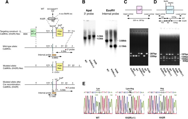
Ca2+/calmodulin-dependent protein kinase IIalpha (CaMKIIalpha) is an essential mediator of activity-dependent synaptic plasticity that possesses multiple protein functions. So far, the autophosphorylation site-mutant mice targeted at T286 and at T305/306 have demonstrated the importance of the autonomous activity and Ca2+/calmodulin-binding capacity of CaMKIIalpha, respectively, in the induction of long-term potentiation (LTP) and hippocampus-dependent learning. However, kinase activity of CaMKIIalpha, the most essential enzymatic function, has not been genetically dissected yet. Here, we generated a novel CaMKIIalpha knock-in mouse that completely lacks its kinase activity by introducing K42R mutation and examined the effects on hippocampal synaptic plasticity and behavioral learning. In homozygous CaMKIIalpha (K42R) mice, kinase activity was reduced to the same level as in CaMKIIalpha-null mice, whereas CaMKII protein expression was well preserved. Tetanic stimulation failed to induce not only LTP but also sustained dendritic spine enlargement, a structural basis for LTP, at the Schaffer collateral-CA1 synapse, whereas activity-dependent postsynaptic translocation of CaMKIIalpha was preserved. In addition, CaMKIIalpha (K42R) mice showed a severe impairment in inhibitory avoidance learning, a form of memory that is dependent on the hippocampus. These results demonstrate that kinase activity of CaMKIIalpha is a common critical gate controlling structural, functional, and behavioral expression of synaptic memory.


Figures

Figure 1.
Figure 1.
Generation of the CaMKIIα (K42R) knock-in mouse. A, K42R mutation was introduced by replacing a nucleotide from AAG to AGG (* on top) within exon 2 of the CaMKIIα gene, which simultaneously generated a new restriction site for BstNI. The homologously recombined ES clones after Cre recombination [CaMKIIα(K42R), bottom] were microinjected into eight-cell embryos to generate the K42R mouse. A, ApaI; BSK, pBluescript SK; EV, EcoRV; H, HindIII; K, KpnI; N, NotI; X, XbaI. B, Southern blot analyses of mouse genomic DNA to confirm homologous recombination. Mouse genomic DNA extracted from tails was digested by ApaI and hybridized with a 3′ external probe, or by EcoRV and with an internal probe. The positions of the probes are indicated in A. K42R, Homozygous K42R mouse; K42R(+/−), heterozygous K42R mouse; WT, wild-type mouse. C, Genotyping of the mouse by PCR amplifying the loxP insertion site to detect a 67 bp size shift. D, Detection of a new BstNI site (*) generated by nucleotide replacement for K42R mutation. The amplified PCR products containing the mutation site were digested by BstNI. E, Direct demonstration of the nucleotide replacement by sequencing the purified PCR products generated as in D.
Figure 2.
Figure 2.
CaMKII isoform expression and CaMKII kinase activity in the CaMKIIα (K42R) knock-in mouse. A, In situ hybridization showing the CaMKII isoform (CaMKIIα, β, γ, and δ) mRNA expression in adult brain sagittal sections (left) and hippocampal coronal sections (right) from wild-type (WT) and homozygous (K42R) mice. CA1–3, Subfield CA1–3 of Ammon's horn; Cb, cerebellum; CP, caudate–putamen; Cx, cerebral cortex; DG, dentate gyrus; Hi, hippocampal formation; MO, medulla oblongata; OB, olfactory bulb; Th, thalamus. B, Immunoblot analyses showing the CaMKII isoform protein levels and the phospho-T286 level of CaMKIIα (P-CaMKIIα) in forebrain and cerebellar homogenates. See also Table 1. C, CaMKII kinase activity in forebrain and cerebellar homogenates from wild-type (WT), heterozygous [K42R(+/−)], and homozygous (K42R) mice. Open columns, Total activity; filled columns, Ca2+/calmodulin-independent autonomous activity. Error bars indicate SEM. ***p < 0.0001, *p < 0.05, ANOVA followed by Fisher's PLSD test (n = 5). See also Table 2. D, CaMKII kinase activity in heterozygous CaMKIIα knock-out mice [CaMKIIα Δ(+/−)] for comparison. **p < 0.01, t test (n = 4). See also Table 3. The CaMKIIα protein level in heterozygous knock-out mice was 39.8 ± 5.3% (p < 0.001, forebrain) and 49.5 ± 15.3% (p < 0.05, cerebellum) of the wild-type level (one-group t test, n = 5).
Figure 3.
Figure 3.
Postsynaptic dynamics of CaMKIIα in CaMKIIα (K42R) neurons. A, FRAP analysis of wild-type and K42R-mutated CaMKIIα tagged with GFP and expressed in hippocampal neurons with respective genotypes. Scale bar, 3 μm. B, FRAP analysis revealed a reduced time constant of K42R CaMKIIα with enhanced fluorescence recovery at the initial three time points (30, 60, and 90 s after bleaching). *p < 0.05, t test. Open circles, Wild type, n = 17 cells; filled circles, K42R, n = 17; three independent preparations from each genotype. Error bars indicate SEM. C, Fluorescence recovery of GFP-Homer1c (wild type, n = 6 cells; K42R, n = 6; one independent preparation from each genotype). D, Local dendritic stimulation by caged glutamate revealed faster translocation of K42R CaMKIIα. The arrows indicate the CaMKIIα clusters analyzed in E. Scale bar, 5 μm. E, Time course of CaMKIIα accumulation induced by glutamate uncaging. Time constants of CaMKIIα accumulation were estimated by exponential fittings. Smaller time constants were observed in homozygous K42R neurons than in wild-type neurons. F, A plot of average time constant against relative fluorescence intensity within dendrites. Open circles, Wild type, n = 15 cells; filled circles, K42R, n = 15; three independent preparations from each genotype.
Figure 4.
Figure 4.
Impaired spine structural plasticity in CaMKIIα (K42R) neurons. A, Extracellular stimulation (100 Hz; 1 s) of GFP-expressing CA1 pyramidal neuronal dendrites in organotypic hippocampal slices from wild-type or homozygous K42R mice. The bottom time-lapse images show structural change of spines (arrows in top images) after electrical stimulation. Spine volume increase was either undetectable (arrowhead) or transient (asterisk) in a K42R neuron. Scale bars, 2 μm. B, Quantification of total fluorescence intensity of spines before and after tetanic stimulation at t = 0. Total fluorescence intensity was normalized by the average of fluorescence intensities before tetanic stimulation. Activity-dependent spine enlargement was severely impaired in K42R neurons. Open circles, Wild type, n = 85 spines (6 slice preparations); filled circles, K42R, n = 73 (9 slice preparations). Error bars indicate SEM. C, Quantification of total fluorescence intensity in spines classified by their immediate response after tetanic stimulation. Spines with a more than twofold increase of total fluorescence intensity at t = 1 min were classified as response (+) (circles), and the other spines were classified as response (−) (triangles) [wild-type response (+), n = 14 spines; K42R response (+), n = 4; wild-type response (−), n = 71; K42R response (−), n = 69].
Figure 5.
Figure 5.
LTP was severely impaired in CaMKIIα (K42R) mice. A, The input–output relationships of AMPAR-mediated EPSPs of wild-type (open circles; n = 12 slices) and homozygous K42R slices (filled circles; n = 10). Error bars indicate SEM. B, AMPAR-mediated (downward traces) and NMDAR-mediated EPSCs (upward traces) in wild-type and homozygous K42R neurons. C, PPF recorded as a function of the interstimulus intervals in the presence of 25 μm d-APV (wild type, n = 8 slices; K42R, n = 8). D, PTP induced by a train of high-frequency stimuli (100 Hz; 1 s) in the presence of 50 μm d-APV (wild type, n = 10 slices; K42R, n = 8). E, The time course of LTP induced by single tetanic stimulation (100 Hz; 1 s). Representative traces (averaged 10 consecutive responses) in the inset are EPSPs obtained at the times indicated by the numbers in the graph (wild type, n = 10 slices; K42R, n = 10). Robust LTP was induced in wild-type slices, whereas no LTP was observed in K42R slices.
Figure 6.
Figure 6.
Inhibitory avoidance learning was severely impaired in CaMKIIα (K42R) mice. A, Wild-type mice showed increased step-through latency compared with training latency, when tested 24 h after one-trial training in the inhibitory avoidance task. Cutoff latency was set at 300 s. Bars represent median. **p < 0.01, Wilcoxon's signed rank test (n = 14). B, Homozygous K42R mice showed no difference between training and 24 h latencies, indicating that avoidance memory was severely impaired in K42R mice (n = 15). Note that shorter training latency in K42R mice (B, left) than in wild-type mice (A, left) reflected hyperactivity in K42R mice (p < 0.01, Mann–Whitney test). C, Short-term memory was tested 40 min after training. Wild-type mice showed increased latency after 40 min, compared with training latency. **p < 0.01, Wilcoxon's signed rank test (n = 11). D, Homozygous K42R mice showed no difference between training and 40 min latencies, indicating that short-term memory was also severely impaired in K42R mice (n = 12). E, Multitrial training was performed to examine immediate learning in homozygous K42R mice. K42R mice that had been subjected to one-trial training and tested 24 h memory in B were retrained after an interval of 5–15 d. The latencies in the first trial (before the first shock) and the second trial (after the first shock) were not different, indicating that immediate learning after a single trial was impaired in K42R mice (n = 14). F, The number of trials necessary for homozygous K42R mice to show immediate avoidance in multitrial training. Training trials were repeated until the mice stayed in the light side for >120 s in a single trial. G, When tested 24 h after multitrial training, homozygous K42R mice showed increased step-through latency compared with the first training latency (E, left) (p < 0.05, Wilcoxon's signed rank test). Note that the 24 h latency after multitrial training in K42R mice (G) was significantly shorter than the 24 h latency after single-trial training in wild-type mice (A, right) (p < 0.01, Mann–Whitney test).
Figure 7.
Figure 7.
Schematic illustration showing that kinase activity of CaMKIIα is essential for structural, functional, and behavioral expression of synaptic memory. Strong synaptic activation induces activation of CaMKII by binding of Ca2+/calmodulin (Ca2+/CaM) and the following postsynaptic translocation of CaMKII. Activated CaMKII undergoes T286 autophosphorylation to generate the autonomous activity and phosphorylates substrate proteins to initiate dendritic spine enlargement and LTP. Such synaptic plasticity seems to be a basis for behavioral expression of learning and memory in vivo. In the kinase-dead CaMKIIα (K42R) knock-in mouse, mutated CaMKIIα did undergo postsynaptic translocation on synaptic activation, and T286 autophosphorylation could occur to some extent by adjacent intact CaMKIIβ within a CaMKII holoenzyme. However, mutated CaMKIIα cannot phosphorylate substrate proteins, and thus, both prolonged dendritic spine enlargement and LTP were not induced. Hippocampus-dependent learning was also impaired in the CaMKIIα (K42R) mouse. These results demonstrate an indispensable role of CaMKIIα kinase activity in dendritic spine enlargement, LTP, and learning.

Similar articles

Cited by

References

    1. Bliss TV, Collingridge GL. A synaptic model of memory: long-term potentiation in the hippocampus. Nature. 1993;361:31–39. - PubMed
    1. Bongsebandhu-phubhakdi S, Manabe T. The neuropeptide nociceptin is a synaptically released endogenous inhibitor of hippocampal long-term potentiation. J Neurosci. 2007;27:4850–4858. - PMC - PubMed
    1. Cammarota M, Bevilaqua LR, Rossato JI, Lima RH, Medina JH, Izquierdo I. Parallel memory processing by the CA1 region of the dorsal hippocampus and the basolateral amygdala. Proc Natl Acad Sci U S A. 2008;105:10279–10284. - PMC - PubMed
    1. Chapman PF, Frenguelli BG, Smith A, Chen CM, Silva AJ. The α-Ca2+/calmodulin kinase II: a bidirectional modulator of presynaptic plasticity. Neuron. 1995;14:591–597. - PubMed
    1. Colbran RJ, Brown AM. Calcium/calmodulin-dependent protein kinase II and synaptic plasticity. Curr Opin Neurobiol. 2004;14:318–327. - PubMed

Publication types

MeSH terms

Substances

LinkOut - more resources