. 2009 Aug;102(2):1017-25.
doi: 10.1152/jn.00100.2009. Epub 2009 Jun 10.

Integration of the reflex pharyngeal swallow into rhythmic oral activity in a neurologically intact pig model

Affiliations

Integration of the reflex pharyngeal swallow into rhythmic oral activity in a neurologically intact pig model

Rebecca Z German et al. J Neurophysiol. 2009 Aug.

Abstract

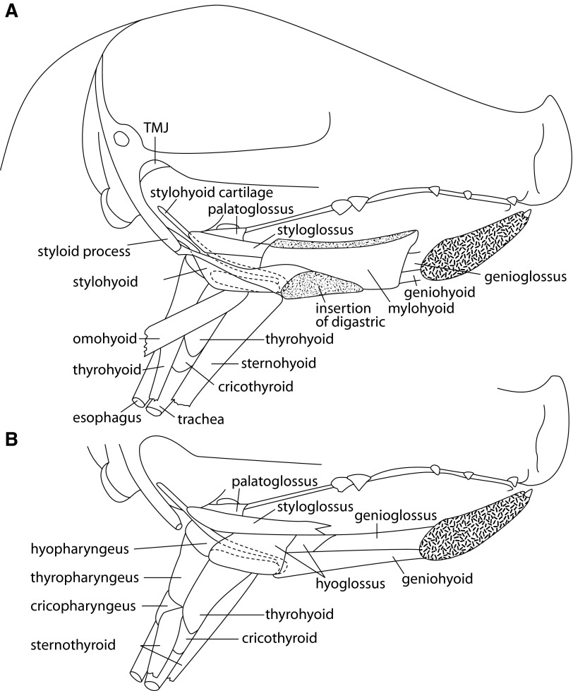
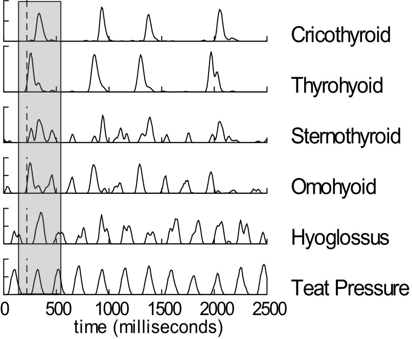
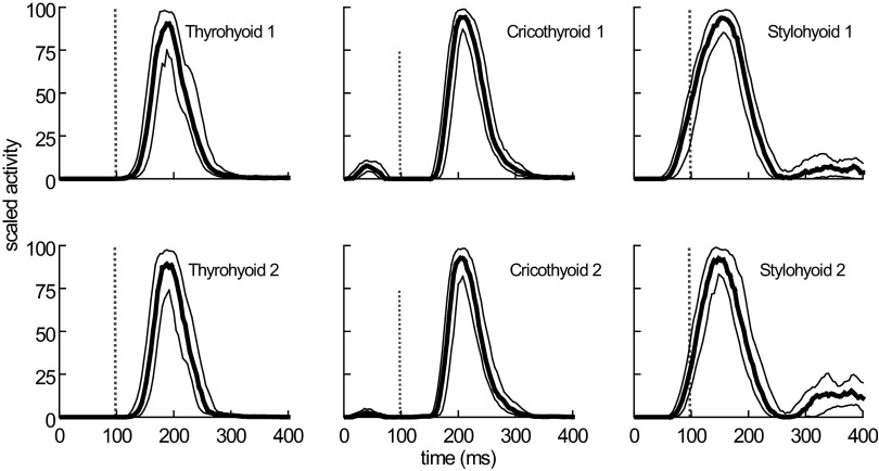
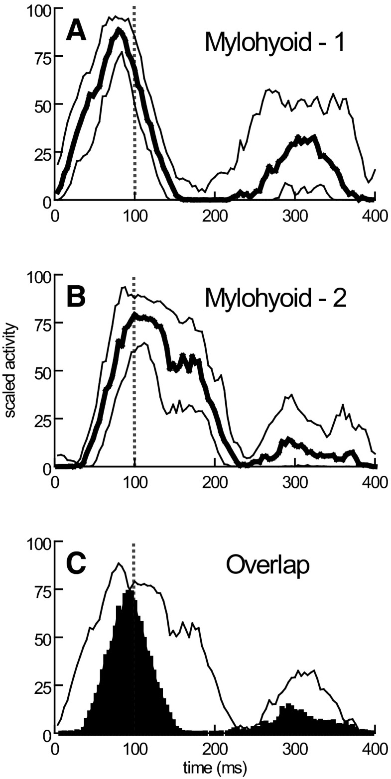
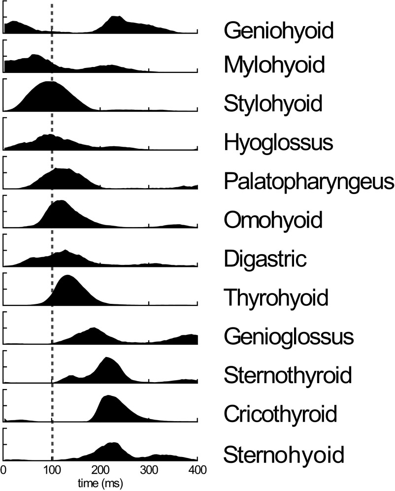
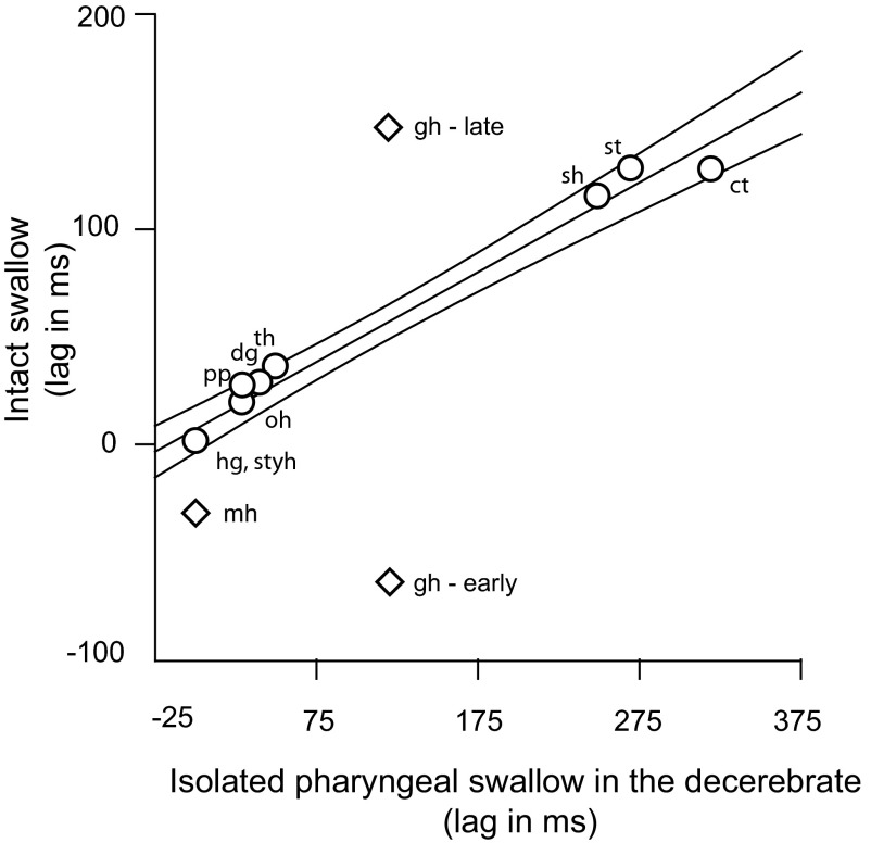
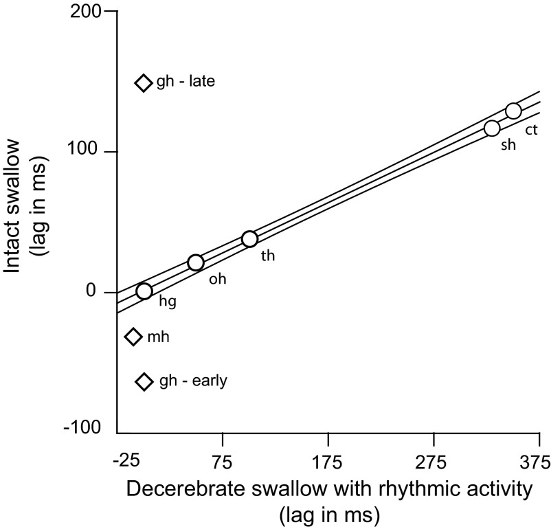
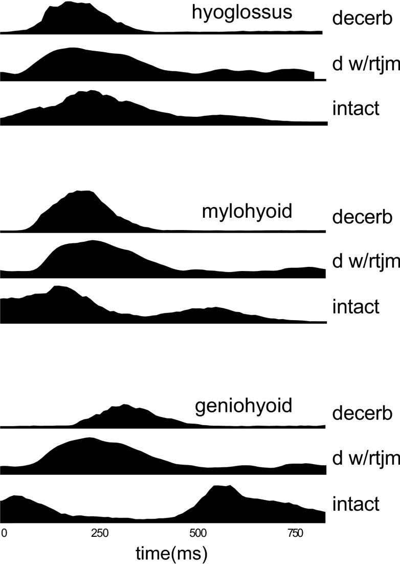
Mammalian swallowing involves the coordinated and sequential activity of many oropharyngeal muscles. Using synchronous electromyography (EMG) and videofluorography, we recorded the pattern of EMG activity for 12 muscles during swallowing in neurologically intact suckling pigs. We tested the hypothesis that this EMG pattern corresponded to the established pattern of activity for the isolated, reflexive pharyngeal swallow of the decerebrate infant pig. The EMG activity associated with the normal swallow of the intact animal had two components: a staggered pattern of single EMG bursts that were prominent in the stylohyoid, thyrohyoid, cricothyroid, and omohyoid muscles and double bursts of activity in some muscles, including geniohyoid and genioglossus, with the same underlying periodicity as suckling. Most of the staggered activity pattern, a linear sequence of progressively delayed activities in different muscles, was not statistically different from that previously found in the reflexive pharyngeal swallow of the decerebrate. However, not all components of the linear sequence of the reflexive swallow were inserted unchanged into the intact swallow. Some components appeared to be delayed or advanced, bringing them into phase with the underlying rhythmic activity. The difference between swallows of intact and of decerebrate animals was not solely due to the presence of rhythmic activity in the former. The timing of some EMG activities in intact animals also differed from the same activities in the few decerebrates that exhibited rhythmic tongue and jaw activity. These results suggest cerebral function influences the EMG pattern of the pharyngeal swallow, which has traditionally been considered a purely reflex pattern.


Figures

FIG. 1.
FIG. 1.
Sagittal section of head of a young pig with mandible removed to show underlying muscles at superficial (A) and deeper (B) levels. TMJ, tempromandibular joint. ···, the hyoid bone.
FIG. 2.
FIG. 2.
Electromyographic (EMG) activity and teat pressures during sucking and suck/swallow cycles in the intact infant pig. Some muscles are activated mainly during swallow cycles, whereas others are activated with every suck cycle. Amplitude is relative to signal maximum over an individual feeding sequence. A total of 12 suck cycles and 4 swallow cycles are shown. Grey bar indicates swallow cycle.
FIG. 3.
FIG. 3.
EMG activity, during successive swallows, recorded simultaneously by duplicate electrodes in 3 muscles. The EMG signals have been processed and the median, upper quartile, and lower quartile profiles of activity derived. The small separation between the quartile profiles is an indication of the low temporal variability of the signals recorded in these 3 muscles.
FIG. 4.
FIG. 4.
Mylohyoid EMG activity recorded by duplicate EMG patch electrodes located at 2 different sites (A and B) on the muscle. The median and quartile profiles of activity indicate both large measures of temporal variability at each site and differences between the timing of signals detected at the 2 sites. The 2 median profiles from A and B are shown in C with their temporal overlap shown in black. The common (black) area is expressed as a percentage of the total area of the 2 median profiles to obtain a measure of the similarity of timing of EMG signals at 2 sites in the same muscle.
FIG. 5.
FIG. 5.
The median profiles of activity during swallowing, across all muscles/animals yielding clean records. ···, the timing of the onset of epiglottal flexion. The successive delays of the profiles of EMG activity were subsequently quantified by cross-correlation, using hyoglossus as the standard.
FIG. 6.
FIG. 6.
Plot of the relative timings of muscle activities in the swallow of an intact conscious animal and the previously reported activity in the isolated pharyngeal swallow of the decerebrate. ○, muscles used in calculating the regression line and the indicated 95% confidence interval (CI). Only 1 value of geniohyoid timing existed in the isolated, decerebrate swallow, but in the data from the intact swallow, there was an early and late burst. Both bursts, and the mylohyoid burst lie outside the CI for the line.
FIG. 7.
FIG. 7.
Plot of the relative timings of muscle activities in the swallow of an intact conscious animal and the previously reported activity in the swallow of the decerebrate exhibiting rhythmic oral activity. Muscles represented by circles were used in calculating the regression line and the indicated 95% confidence interval. Only 1 value of geniohyoid timing existed in the decerebrate swallow with rhythmic activity, but in the data from the intact swallow, there was an early and late burst. Both bursts and the mylohyoid burst lie outside the CI for the line.
FIG. 8.
FIG. 8.
The relationship between the EMG activities in three key muscles for arrhythmic decerebrates (dec), decerebrates exhibiting rhythmic tongue and jaw movement (dec+rtjm) and conscious intact suckling pigs (intact). The data from each of the 3 sources has been aligned to the profiles of hyoglossus activity using the cross-correlations between the median profiles of hyoglossus EMG activity to mylohyoid and geniohyoid EMG activity. The time scale is the relative 100 units used for each swallow because of the differences in swallow durations among the 3 models. The median profiles of mylohyoid EMG activity suggest a slightly earlier response in the presence of cerebral hemispheres while the median profiles of geniohyoid EMG activity indicate a substantially changed pattern of response.

Similar articles

Cited by

References

    1. Amarasena J, Ootaki S, Yamamura K, Yamada Y. Effect of cortical masticatory area stimulation on swallowing in anesthetized rabbits. Brain Res 965: 222–238, 2003. - PubMed
    1. Basmajian J, Stecko G. A new bipolar electrode for electromyography. J Appl Physiol 17: 849, 1962.
    1. Bremer F Physiologie nerveuse de la mastication chez le chat et le lapin. Arch Int Physiol 21: 309–352, 1923.
    1. Buchthal F, Schmalbruch H. Motor unit of mammalian muscle. Physiol Rev 60: 901–942, 1980. - PubMed
    1. Crompton AW, German RZ, Thexton AJ. Development of the movement of the epiglottis in infant and juvenile pigs. Zoology 111: 339–349, 2008. - PMC - PubMed

Publication types

LinkOut - more resources