. 2009 Aug;297(2):R470-7.
doi: 10.1152/ajpregu.90821.2008. Epub 2009 Jun 10.

Dorsal spinal cord stimulation obtunds the capacity of intrathoracic extracardiac neurons to transduce myocardial ischemia

Affiliations

Dorsal spinal cord stimulation obtunds the capacity of intrathoracic extracardiac neurons to transduce myocardial ischemia

Jeffrey L Ardell et al. Am J Physiol Regul Integr Comp Physiol. 2009 Aug.

Abstract

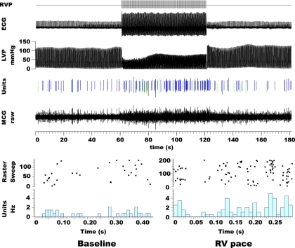
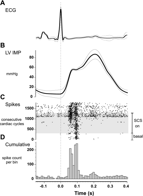
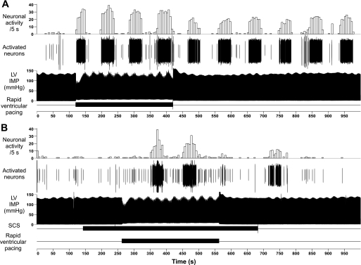
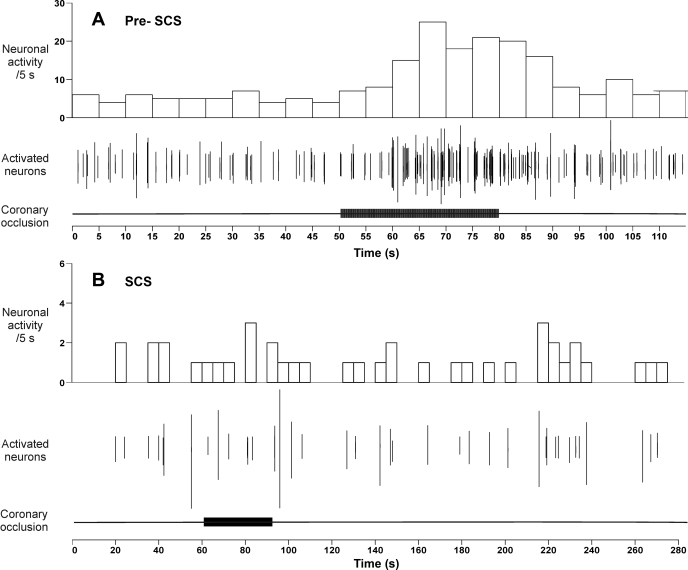
Populations of intrathoracic extracardiac neurons transduce myocardial ischemia, thereby contributing to sympathetic control of regional cardiac indices during such pathology. Our objective was to determine whether electrical neuromodulation using spinal cord stimulation (SCS) modulates such local reflex control. In 10 anesthetized canines, middle cervical ganglion neurons were identified that transduce the ventricular milieu. Their capacity to transduce a global (rapid ventricular pacing) vs. regional (transient regional ischemia) ventricular stress was tested before and during SCS (50 Hz, 0.2 ms duration at 90% MT) applied to the dorsal aspect of the T1 to T4 spinal cord. Rapid ventricular pacing and transient myocardial ischemia both activated cardiac-related middle cervical ganglion neurons. SCS obtunded their capacity to reflexly respond to the regional ventricular ischemia, but not rapid ventricular pacing. In conclusion, spinal cord inputs to the intrathoracic extracardiac nervous system obtund the latter's capacity to transduce regional ventricular ischemia, but not global cardiac stress. Given the substantial body of literature indicating the adverse consequences of excessive adrenergic neuronal excitation on cardiac function, these data delineate the intrathoracic extracardiac nervous system as a potential target for neuromodulation therapy in minimizing such effects.


Figures

Fig. 1.
Fig. 1.
Ventricular pacing modified cardiac-related middle cervical ganglion (MCG) neuronal activity. Top: rapid right ventricular pacing (RVP; initiated throughout the RVP signal on upper trace) induced a fall in left ventricular pressure (LVP) while activating 3 neurons identified from raw MCG neuronal activity (line below) by their unique action potential configurations (green, blue, and black vertical lines). Bottom: raster sweep plots of the action potentials generated by these 3 neurons during consecutive cardiac cycles for 60 s during control states (baseline) and right ventricular pacing (RV pace). Time 0 is defined by QRS onset. Activity increased during pacing, rapidly returning to control values once pacing ceased (cf, unit activity). Timing bars are located below each panel.
Fig. 2.
Fig. 2.
Spinal cord stimulation (SCS) modified the activity generated by MCG neurons associated with left ventricular mechanosensory neurites. Lead II electrocardiogram (ECG) (A) and left ventricular intramyocardial pressure (LV IMP) (B) show QRS averaged values (dark line) recorded during each of 1,792 consecutive cardiac cycles (± 1 SD thin lines). C: dots in the raster sweep plot represent action potentials generated by 4 neurons recorded during the 1,792 cardiac cycles. This included activity recorded 2 min before (basal) and during 9 min of continuous SCS (SCS on = gray shaded area), as well as 4 min following SCS (top of raster plot). The horizontal time 0 was set to the R wave of the ECG. Activity increased as SCS persisted (gray zone in raster plot), including after SCS termination (top of this plot C); increased activity demonstrated less phase locking to the cardiac cycle. D: cumulative index of all action potentials recorded during these 1,792 consecutive cardiac cycles demonstrating that activity was maximal during maximum local isovolumetric dynamics of their receptor fields.
Fig. 3.
Fig. 3.
A: rapid ventricular pacing (horizontal bar below) increased neuronal activity. A maximum of 5 identified middle cervical ganglion neurons were recorded that generated increased (cyclic bursts) activity during pacing, something that persisted for some time after discontinuing pacing. B: 9 min of SCS (started 2 min prior to pacing; horizontal bar below) induced a lesser response to pacing (lowest horizontal bar) and one that extinguished soon after discontinuing SCS. In each panel, from above downward, are: neuronal activity averaged over 5-s sequential intervals, discriminated neuronal activity (activated neurons), and intramyocardial pressure (LV IMP) recorded adjacent to the identified sensory neurite field. The horizontal bar below represents SCS application time (applied 2 min before and continuing for 2 min after pacing).
Fig. 4.
Fig. 4.
Effects of left anterior descending coronary artery occlusion (30-s duration; horizontal bars below each panel) on middle cervical ganglion neuronal activity before (Pre-SCS) (A) and during (B) SCS. The time scales of these 2 records differ in order to emphasize the lack of long-term effects of ischemia after SCS. Neuronal activity and activated neuron traces are as defined in Fig 1.

References

    1. American Physiological Society. Guiding principles for research involving animals and human beings. Am J Physiol Regul Integr Comp Physiol 283: R281–R283, 2002. - PubMed
    1. Andresen MC, Kunze DL, Mendelowitz D. Central nervous system regulation of the heart. In: Basic and Clinical Neurocardiology, edited by Armour JA and Ardell JL. New York: Oxford University Press, 2004, p. 187–219.
    1. Ardell JL Intrathoracic neuronal regulation of cardiac function. In: Basic and Clinical Neurocardiology, edited by Armour JA and Ardell JL. New York: Oxford University Press, 2004, p. 118–152.
    1. Ardell JL, Butler CK, Smith FM, Hopkins DA, Armour JA. Activity of in vivo atrial and ventricular neurons in chronic decentralized canine hearts. Am J Physiol Heart Circ Physiol 260: H713–H721, 1991. - PubMed
    1. Armour JA Synaptic transmission in chronically decentralized middle cervical and stellate ganglia of the dog. Can J Physiol Pharmacol 61: 1149–1155, 1983. - PubMed

Publication types

MeSH terms

LinkOut - more resources