

Speckle Reduction in OCT using Massively-Parallel Detection and Frequency-Domain Ranging
- PMID: 19516630
- PMCID: PMC2704480
- DOI: 10.1364/oe.14.004736
Speckle Reduction in OCT using Massively-Parallel Detection and Frequency-Domain Ranging
Abstract
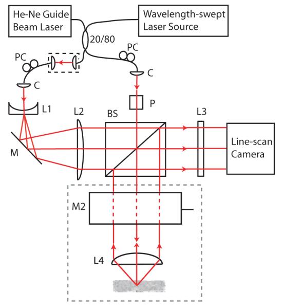
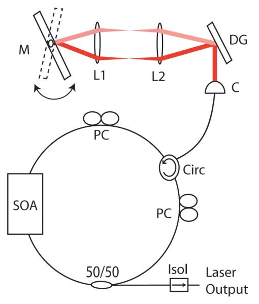
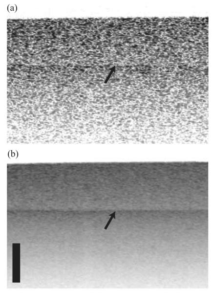
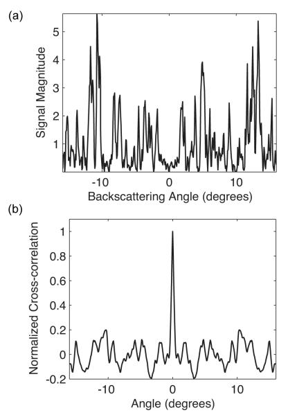
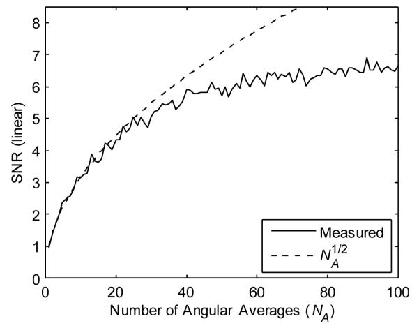
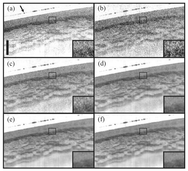
Speckle noise significantly limits the information content provided by coherent optical imaging methods such as optical coherence tomography and its recent derivative, optical frequency-domain imaging (OFDI). In this paper, we demonstrate a novel OFDI system that simultaneously acquires hundreds of angularly resolved images, which can be compounded to reduce speckle noise. The system comprises an InGaAs line-scan camera and an interferometer, configured so that the elements of the detector array simultaneously capture light spanning a backscattering angular range of 32 degrees. On successive read-outs of the array, the wavelength of the laser source was stepped through a range of 130 nm centered at 1295 nm to concurrently generate 400 angle-resolved OFDI images. A theory of angle-resolved OFDI and the design equations of the system are presented. Incoherent averaging of the angle-resolved data is shown to yield substantial speckle reduction (as high as an 8 dB SNR improvement) in images of a tissue phantom and esophageal tissue ex vivo.
Figures
References
-
- Huber R, Wojtkowski M, Fujimoto JG, Jiang JY, Cable AE. “Three-dimensional and C-mode OCT imaging with a compact, frequency swept laser source at 1300 nm,”. Opt. Express. 2005;13:10523–10538. - PubMed
-
- Choma MA, Hsu K, Izatt JA. “Swept source optical coherence tomography using an all-fiber 1300-nm ring laser source,”. J. Biomed. Opt. 2005;10:044009. - PubMed
-
- Yung KM, Lee SL, Schmitt JM. “Phase-domain processing of optical coherence tomography images,”. J. Biomed. Opt. 1999;4:125–136. - PubMed
-
- Pircher M, Gotzinger E, Leitgeb R, Fercher AF, Hitzenberger CK. “Speckle reduction in optical coherence tomography by frequency compounding,”. J. Biomed. Opt. 2003;8:565–569. - PubMed
Grants and funding
LinkOut - more resources
Full Text Sources
Other Literature Sources
Miscellaneous
