. 2009 May;109(4):981-94.
doi: 10.1111/j.1471-4159.2009.06034.x. Epub 2009 Mar 11.

Interaction of catechol and non-catechol substrates with externally or internally facing dopamine transporters

Affiliations

Interaction of catechol and non-catechol substrates with externally or internally facing dopamine transporters

Ying-Jian Liang et al. J Neurochem. 2009 May.

Abstract

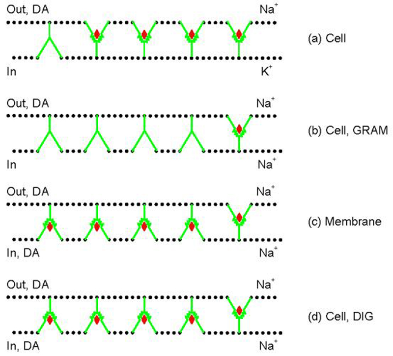
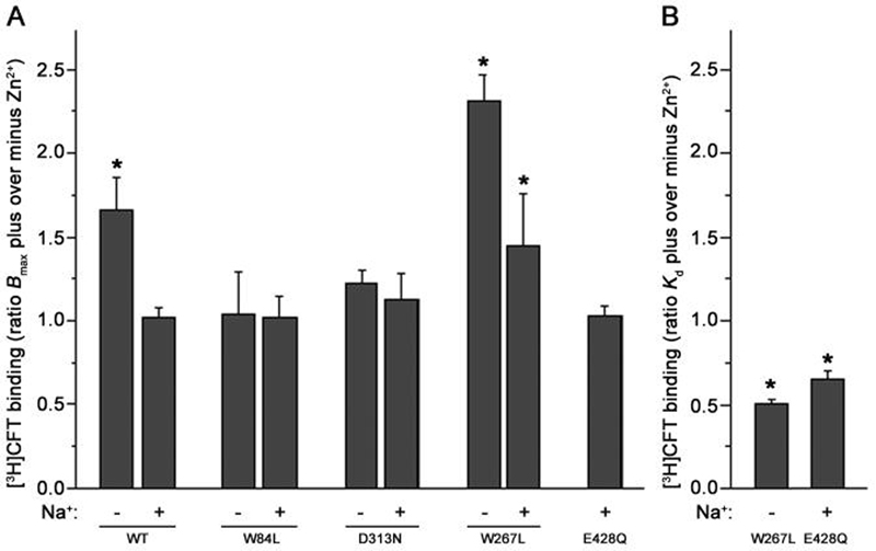
Our previous work suggested that collapsing the Na(+) gradient and membrane potential converts the dopamine (DA) transporter (DAT) to an inward-facing conformation with a different substrate binding profile. Here, DAT expressing human embryonic kidney 293 cells were permeabilized with digitonin, disrupting ion/voltage gradients and allowing passage of DAT substrates. The potency of p-tyramine and other non-catechols (d-amphetamine, beta-phenethylamine, MPP(+)) in inhibiting cocaine analog binding to DAT in digitonin-treated cells was markedly weakened to a level similar to that observed in cell-free membranes. In contrast, the potency of DA and another catechol, norepinephrine, was not significantly changed by the same treatment, whereas epinephrine showed only a modest reduction. These findings suggest that catechol substrates interact symmetrically with both sides of DAT and non-catechol substrates, favoring binding to outward-facing transporter. In the cocaine analog binding assay, the mutant W84L displayed enhanced intrinsic binding affinity for substrates in interacting with both outward- and inward-facing states; D313N showed wild-type-like symmetric binding; but D267L and E428Q showed an apparent improvement in the permeation pathway from the external face towards the substrate site. Thus, the structure of both substrate and transporter play a role in the sidedness and mode of interaction between them.


Figures

Fig. 1
Fig. 1. Cartoon of access model for DA at wild type DAT
(a) In intact cells under physiological condition, most DATs reside in outward-facing state where extracellular DA binding sites are accessible. K+ prevents internally accumulated DA to bind to the few inward-facing transporters; for simplicity internal DA has been omitted from the cartoon here and in Figs. 6 and 7 (Cell Control). (b) Gramicidin (GRAM) treatment causes the collapse of membrane Na+ gradient (high [Na+]o and low [Na+]i), leading to accumulation of the transporter in inward-facing state. Under this condition, the majority of binding sites is unavailable to extracellular DA. Intracellular DA is sparse. (c) In cell-free membranes, the complete disruption of the membrane Na+ gradient causes DAT redistribution similar to that accruing in gramicidin-treated cells. Ambient DA can approach its binding site either externally or internally. (d) Digitionin (DIG) treatment causes a similar change of transporter conformation as in GRAM-treated cells or cell-free membranes, but (in contrast to the situation with GRAM) DA can access from both sides similar to the case for cell-free membranes; DIG holds the cellular milleu similar to that occurring in GRAM-treated cells. Note that all panels illustrate only the initial conformational state for ligand binding. Cartoons are drawn according to those presented for Tyt1 (Quick et al. 2006), another Na+-dependent bacterial transporter for tyrosine operating with the same gated pore mechanism for transport as the DAT.
Fig. 2
Fig. 2. Effect of digitonin treatment on CFT and DA binding
Intact cells (A) and cell-free membranes (B) were pretreated with 15 μM digitonin (DIG +) or vehicle (0.75% ethanol) (−) for 15 min at 21°C, and then subjected to the binding assay with 2-4 nM [3H]CFT and various concentrations of CFT or DA for 15 min at 21°C. *P < 0.05 compared with respective WT within the same treatment group (+ or − DIG) (Student's t test); #P < 0.05 compared with respective value in cell-free membranes (depicted in panel B) for same DAT construct (Student's t test). Note there is no statistically significant difference in CFT Kd between digitonin and vehicle treatment for Cell or Membrane preparations of a given DAT construct (Student's t test, paired where appropriate).
Fig. 3
Fig. 3. Potency of catechol and non-catechol substrates in inhibiting [3H]CFT binding to wild type DAT
Intact cells (Cell) and cell-free membranes (Mem) were pretreated with 15 μM digitonin or 0.75% ethanol vehicle (control) for 15 min at room temperature, and then subjected to the binding assay with 2-4 nM [3H]CFT and various concentrations of DA, norepinephrine (NE), epinephrine (EPI), d-amphetamine (d-AMPH), β-phenethylamine (β-PE), p-tyramine (p-TYR) and MPP+ for 15 min at 21°C. #P < 0.05 compared with control Ki of respective Cell or Mem (Student's t test); *P < 0.05 compared with the Ki for Cell within the same treatment group (control or digitonin) (Student's t test). Chemical structure of each substrate is shown above the chart.
Fig. 4
Fig. 4. Effect of Zn2+ on transporter conformation in cells not treated with digitonin
Intact cells stably expressing WT DAT or its mutants (W84L, D313N, W267L or E428Q) were subjected to the binding assay with 2-4 nM [3H]CFT and various concentrations of CFT with or without 10 μM Zn2+ for 15 min at 21°C. The ratio of the Bmax of [3H]CFT binding in the presence or absence of Zn2+ is shown in A. The ratio of the Kd in Zn2+ treated cells to that in untreated control cells is shown in B only for those changes that were statistically significant. The +Na+ and −Na+ bars indicate the presence or absence of 130 mM NaCl in the binding assay respectively, for details see Materials and Methods. Note that the E428Q mutant displayed too low [3H]CFT binding in the absence of Na+ to be analyzed. *P < 0.05 compared with ratio of unity (one-sample Student's t-test, two-tailed).
Fig. 5
Fig. 5. Effect of Zn2+ on transporter conformation in cells treated with digitonin
Digitonin-permeabilized cells were incubated with 4 nM [3H]CFT and 10 μM Zn2+ in KRH buffer with 130 mM NaCl. Data are expressed as the ratio of [3H]CFT binding (at the same protein level) in the presence over that in the absence of Zn2+. *P < 0.05 compared with WT (one-way ANOVA followed by Dunnett multiple comparisons test).
Fig. 6
Fig. 6. Cartoon of binding model for DA at wild-type and mutant DATs
Conformational distributions between outward and inward states are drawn qualitatively based on the Zn2+ experiments. WT: DATs in control cells are mostly outward-facing; in digitonin (DIG)-treated cells and membranes (either with or without DIG treatment), DAT is mostly inward-facing. DA binds DAT with similar potency in both situations. W84L: Conformational preference is the same as that for WT, except for a somewhat reduced conversion to inward-facing transporters by DIG. DA is able to reach both external and internal binding sites as WT. However, the intrinsic binding affinity is enhanced at both sides (indicated by a better fit of the red diamond). D313N: Conformational preference and DA binding pattern are similar as for WT except for a somewhat reduced conversion to inward-facing transporters by DIG. D267L: In control cells, less transporter is facing outward compared with that in WT, but DA can access its binding site on DAT with greater ease (indicated by blue arrow) resulting in a higher apparent affinity compared with that at WT. In DIG-treated cells and in membranes, DAT is mostly inward-facing and the DA-DAT interaction is similar to that for WT; by analogy with the comparable case in Fig. 7, the enhanced access to outward-facing DAT at the right hand has been removed (see Fig. 7 legend). E428Q: In cells, DATs are mostly outward facing as for WT; in membranes, the reversed is true as is the case for WT. DA has free access to external and internal binding sites. However, outward-facing DAT displays higher apparent affinity for DA compared to WT because of easier access of DA to its binding site (blue arrow); by analogy with the comparable case in Fig. 7, the enhanced access to outward-facing DAT at the right hand has been removed (see Fig. 7 legend). For each cell type, the number in the left parentheses is the DA Ki in control cells; the first number in the right parentheses is the DA Ki in cells treated with DIG, the second number is the mean of DA Ki values from membranes treated with or without DIG. Changes in DA recognition or access in the mutants are drawn only when the data indicated statistically significant differences with WT.
Fig. 7
Fig. 7. Cartoon of binding model for p-tyramine at wild-type and mutant DATs
DAT conformational distribution in control cells, digitonin (DIG)-permeabilized cells and in membranes is the same as in Figure 5. p-Tyramine only binds to outward-facing transporters. For control cells, W84L is prone to have a higher affinity for DA compared with WT (indicated by better fit of violet diamond). W267L and E428Q have a higher apparent affinity by easier access (indicated by blue arrow). For DIG-permeabilized cells and for membranes, p-tyramine binds to WT and the mutants similarly, except in the case of W84L that displays a higher affinity (in analogy with control cells and with DA in Fig. 6 in the comparable case). Depolarization combined with disruption of the transmembrane ion gradient (panels on the right) is speculated to remove the enhanced access for W267L and E428Q. For each cell type, the number in the left parentheses is the p-tyramine Ki in control cells; the first number in the right parentheses is the p-tyramine Ki in cell treated with DIG, the second number is the mean of p-tyramine Ki values from membranes treated with or without DIG.

Similar articles

Cited by

References

    1. Amara SG, Sonders MS. Neurotransmitter transporters as molecular targets for addictive drugs. Drug Alcohol Depend. 1998;51:87–96. - PubMed
    1. Amejdki-Chab N, Costentin J, Bonnet JJ. Kinetic analysis of the chloride dependence of the neuronal uptake of dopamine and effect of anions on the ability of substrates to compete with the binding of the dopamine uptake inhibitor GBR 12783. J. Neurochem. 1992;58:793–800. - PubMed
    1. Androutsellis-Theotokis A, Rudnick G. Accessibility and conformational coupling in serotonin transporter predicted internal domains. J. Neurosci. 2002;22:8370–8378. - PMC - PubMed
    1. Appell M, Berfield JL, Wang LC, Dunn WJ, III, Chen N, Reith ME. Structure-activity relationships for substrate recognition by the human dopamine transporter. Biochem. Pharmacol. 2004;67:293–302. - PubMed
    1. Bannon MJ. The dopamine transporter: role in neurotoxicity and human disease. Toxicol. Appl. Pharmacol. 2005;204:355–360. - PubMed

Publication types