Review
. 2009;11(3):224.
doi: 10.1186/ar2592. Epub 2009 May 19.

Cartilage homeostasis in health and rheumatic diseases

Affiliations
Review

Cartilage homeostasis in health and rheumatic diseases

Mary B Goldring et al. Arthritis Res Ther. 2009.

Abstract

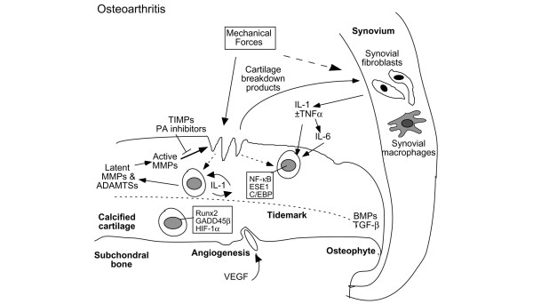
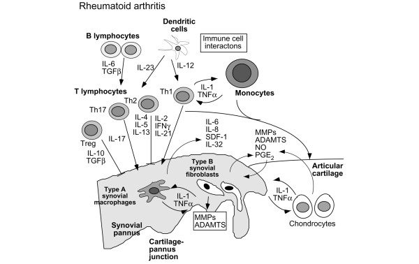
As the cellular component of articular cartilage, chondrocytes are responsible for maintaining in a low-turnover state the unique composition and organization of the matrix that was determined during embryonic and postnatal development. In joint diseases, cartilage homeostasis is disrupted by mechanisms that are driven by combinations of biological mediators that vary according to the disease process, including contributions from other joint tissues. In osteoarthritis (OA), biomechanical stimuli predominate with up-regulation of both catabolic and anabolic cytokines and recapitulation of developmental phenotypes, whereas in rheumatoid arthritis (RA), inflammation and catabolism drive cartilage loss. In vitro studies in chondrocytes have elucidated signaling pathways and transcription factors that orchestrate specific functions that promote cartilage damage in both OA and RA. Thus, understanding how the adult articular chondrocyte functions within its unique environment will aid in the development of rational strategies to protect cartilage from damage resulting from joint disease. This review will cover current knowledge about the specific cellular and biochemical mechanisms that regulate cartilage homeostasis and pathology.


Figures

Figure 1
Figure 1
Cellular interactions in cartilage destruction in osteoarthritis. This scheme represents the destruction of the cartilage due to mechanical loading and biological factors. The induction of stress-induced intracellular signals, catabolic cytokines, including interleukin-1 (IL-1) and tumor necrosis factor-alpha (TNF-α), chemokines, and other inflammatory mediators produced by synovial cells and chondrocytes results in the upregulation of cartilage-degrading enzymes of the matrix metalloproteinase (MMP) and ADAMTS families. Matrix degradation products can feedback regulate these cellular events. Anabolic factors, including bone morphogenetic proteins (BMPs) and transforming growth factor-beta (TGF-β), may also be upregulated and participate in osteophyte formation. In addition to matrix loss, evidence of earlier changes, such as chondrocyte proliferation and hypertrophy, increased cartilage calcification with tidemark advancement, and microfractures with angiogenesis from the subchondral bone possibly mediated by vascular endothelial growth factor (VEGF) can be observed in late osteoarthritis samples obtained from patients after total joint replacement. ADAMTS, a disintegrin and metalloproteinase with thrombospondin-1 domains; C/EBP, CCAAT enhancer-binding protein; ESE1, epithelial-specific ETS; ETS, E26 transformation specific; GADD45β, growth arrest and DNA damage 45 beta; HIF-1α, hypoxia-inducible factor-1-alpha; NF-κB, nuclear factor-kappa-B; PA, plasminogen activator; TIMPs, tissue inhibitors of metalloproteinases.
Figure 2
Figure 2
Cellular interactions in cartilage destruction in rheumatoid arthritis. This scheme represents the progressive destruction of the cartilage associated with the invading synovial pannus in rheumatoid arthritis. As a result of immune cell interactions involving T and B lymphocytes, monocytes/macrophages, and dendritic cells, a number of different cytokines are produced in the synovium due to the influx of inflammatory cells from the circulation and synovial cell hyperplasia. The induction of proinflammatory cytokines produced primarily in the synovium, but also by chondrocytes, results in the upregulation of cartilage-degrading enzymes at the cartilage-pannus junction. Chemokines, nitric oxide (NO), and prostaglandins (PGE2) also contribute to the inflammation and tissue catabolism. ADAMTS, a disintegrin and metalloproteinase with thrombospondin-1 domains; IFN-γ, interferon-gamma; IL, interleukin; MMP, matrix metalloproteinase; SDF-1, stromal derived factor 1; TGF-β, transforming growth factor-beta; TNF-α, tumor necrosis factor-alpha; Treg, regulatory T (cell).

References

    1. Goldring MB, Goldring SR. Osteoarthritis. J Cell Physiol. 2007;213:626–634. - PubMed
    1. Dayer JM. The process of identifying and understanding cytokines: from basic studies to treating rheumatic diseases. Best Pract Res Clin Rheumatol. 2004;18:31–45. - PubMed
    1. Loo FA van de, Geurts J, Berg WB van den. Gene therapy works in animal models of rheumatoid arthritis...so what! Curr Rheumatol Rep. 2006;8:386–393. - PubMed
    1. Berg WB van den. Lessons from animal models of osteoarthritis. Curr Rheumatol Rep. 2008;10:26–29. - PubMed
    1. Poole AR. Cartilage in health and disease. In: Koopman WS, editor. Arthritis and Allied Conditions: A Textbook of Rheumatology. 15. Philadelphia: Lippincott, Williams, and Wilkins; 2005. pp. 223–269.

Publication types