Review
. 2009 Oct;30(4):429-441.
doi: 10.1016/j.yfrne.2009.05.007. Epub 2009 Jun 9.

Dynamic limbic networks and social diversity in vertebrates: from neural context to neuromodulatory patterning

Affiliations
Review

Dynamic limbic networks and social diversity in vertebrates: from neural context to neuromodulatory patterning

James L Goodson et al. Front Neuroendocrinol. 2009 Oct.

Abstract

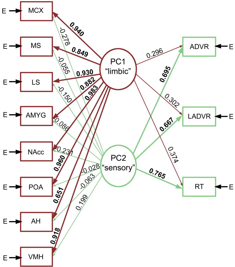
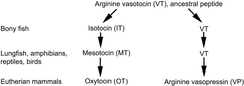
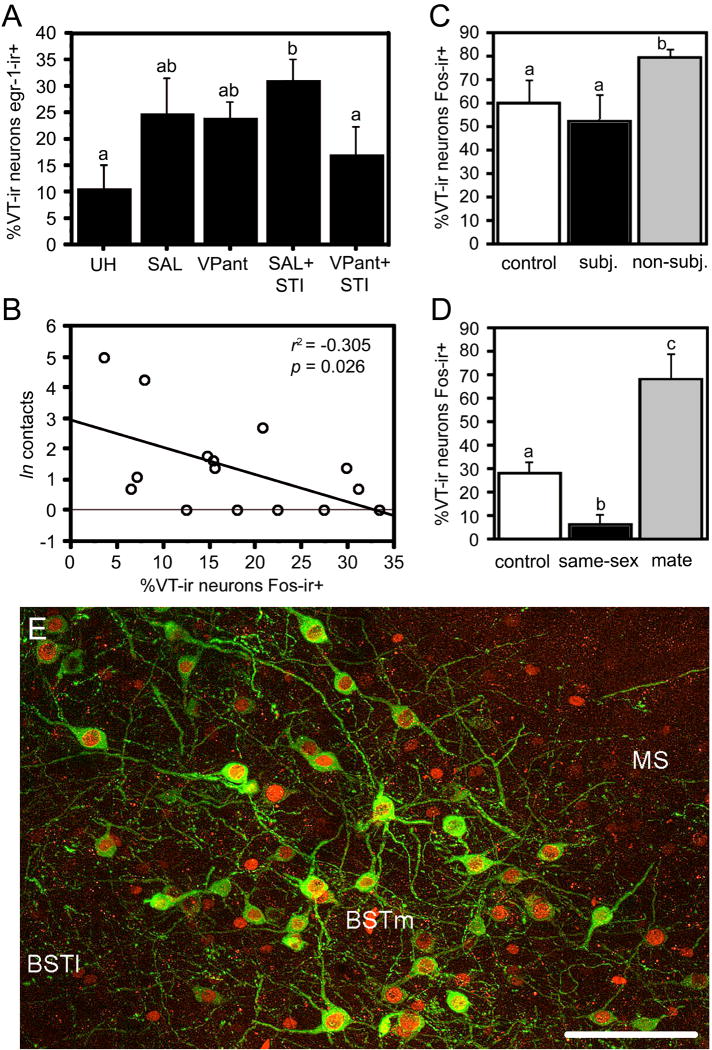
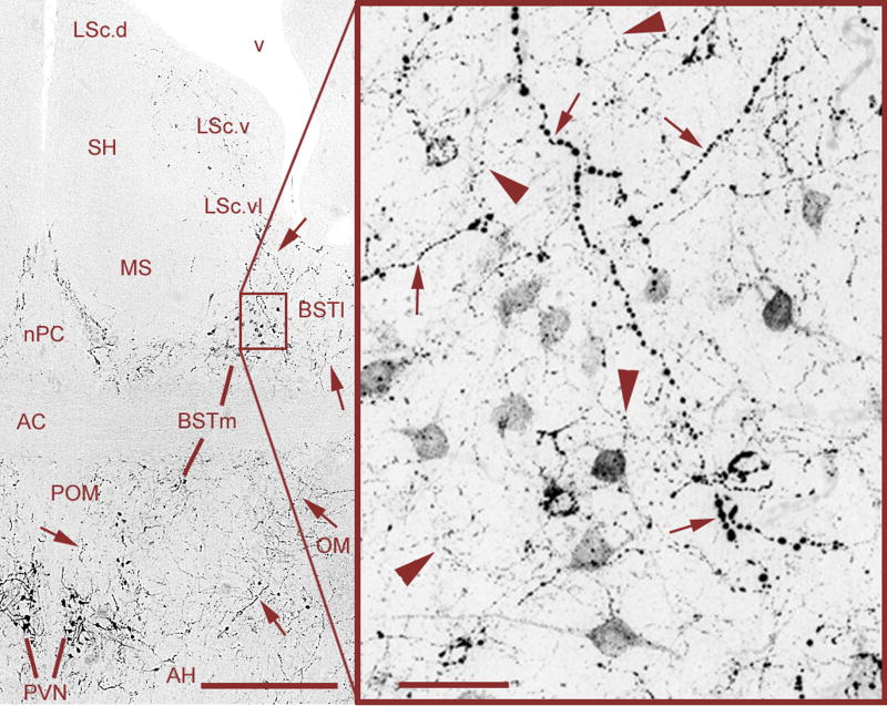
Vertebrate animals exhibit a spectacular diversity of social behaviors, yet a variety of basic social behavior processes are essential to all species. These include social signaling; discrimination of conspecifics and sexual partners; appetitive and consummatory sexual behaviors; aggression and dominance behaviors; and parental behaviors (the latter with rare exceptions). These behaviors are of fundamental importance and are regulated by an evolutionarily conserved, core social behavior network (SBN) of the limbic forebrain and midbrain. The SBN encodes social information in a highly dynamic, distributed manner, such that behavior is most strongly linked to the pattern of neural activity across the SBN, not the activity of single loci. Thus, shifts in the relative weighting of activity across SBN nodes can conceivably produce almost limitless variation in behavior, including diversity across species (as weighting is modified through evolution), across behavioral contexts (as weights change temporally) and across behavioral phenotypes (as weighting is specified through heritable and developmental processes). Individual neural loci may also express diverse relationships to behavior, depending upon temporal variations in their functional connectivity to other brain regions ("neural context"). We here review the basic properties of the SBN and show how behavioral variation relates to functional connectivity of the network, and discuss ways in which neuroendocrine factors adjust network activity to produce behavioral diversity. In addition to the actions of steroid hormones on SBN state, we examine the temporally plastic and evolutionarily labile properties of the nonapeptides (the vasopressin- and oxytocin-like neuropeptides), and show how variations in nonapeptide signaling within the SBN serve to promote behavioral diversity across social contexts, seasons, phenotypes and species. Although this diversity is daunting in its complexity, the search for common "organizing principles" has become increasingly fruitful. We focus on multiple aspects of behavior, including sexual behavior, aggression and affiliation, and in each of these areas, we show how broadly relevant insights have been obtained through the examination of behavioral diversity in a wide range of vertebrate taxa.


Figures

Fig. 1
Fig. 1
Aggressive experience in male anoles (repeated exposure to an aggressive video) organizes multiple functionally connected neural networks, including a sensory network and a limbic-hypothalamic network that included all forebrain components of the SBN. Shown here is a path diagram based upon the principle component (PC) structure of males exposed to videos. Arrow E represents variances contributed by factors other than the linear combination of the latent variables derived from the PCs. The thickness of the lines and the size of the numerals (PC loadings) indicate the strength of the relationships. Modified from [118]. Abbreviations: ADVR, anterior dorsal ventricular ridge; AH, anterior hypothalamus; AMYG, amygdala; LADVR, lateral ADVR; LS, lateral septum; MCX, medial cortex; MS, medial septum; NAcc, nucleus accumbens; POA, preoptic area; RT, nucleus rotundus; VMH, ventromedial hypothalamus.
Fig. 2
Fig. 2
Evolutionary history of the vertebrate nine-amino acid “nonapeptides.” Arginine vasotocin (VT) is the ancestral peptide form. Separate clades giving rise to mammalian vasopressin (VP) and oxytocin (OT) originated with a duplication of the VT gene in early jawed vertebrates. Each change in form (e.g., from isotocin, IT, to mesotocin, MT) represents a single amino acid substitution, but OT differs from VT at only one position. Only major features of the family are shown, and some species show unique peptide forms. For reviews, see [2,70,71].
Fig. 3
Fig. 3
Functional profiles of VT cell groups in the PVN and BSTm of songbirds, showing that these cell groups respond to negative and positive stimuli, respectively. (A) Colocalization of VT and egr-1 (Zenk) in male song sparrows housed on semi-natural territories (field-based flight cages) following control conditions (unhandled; UH); capture and restraint for i.c.v. infusion of saline (SAL) or a VP antagonist (VPant); or infusions followed by simulated territorial intrusions (STI; presentation of a caged decoy and playback of song). Total n = 21. Modified from [54]. Although VT-Zenk colocalization increases following STI, findings in male song sparrows (B) show that VT-Fos colocalization in the PVN correlates negatively with an index of aggressive response (“contacts;” n = 16; J.L. Goodson, A.K. Evans and K.K. Soma, unpubl. obs.). Whereas VT-Fos colocalization in the BSTm was unaffected by STI (not shown), it does increase in zebra finches following aggressive competition for mates (C), but not in subjects that are intensely subjugated (subj.) and not allowed to court (total n = 15; sexes pooled). This pattern of results for the BSTm reflects a sensitivity to stimulus valence. For instance, whereas exposure to a same-sex conspecific produces a significant increase in VT-Fos colocalization in the BSTm of zebra finches, which are gregarious (see section 5.4), same-sex stimuli produce a decrease in VT-Fos colocalization in the BSTm of territorial violet-eared waxbills (D). Violet-eared waxbills nonetheless exhibit robust increases in colocalization following exposure to a positive social stimulus (the subject’s pair bond partner, or “mate;” also in panel D; total n = 16; sexes pooled). Modified from [62]. Bar graphs present means ± SEM. Different letters above the error bars denote significant group differences. (E) Representative labeling for VT (Alexa Fluor 488; green) and Fos (Alexa Fluor 594; red) in the BSTm of a male zebra finch following exposure to a female. Abbreviations: BSTl, lateral bed nucleus of the stria terminalis; MS, medial septum. Scale bar = 50 μm. Modified from [61].
Fig. 4
Fig. 4
Mixed VT signals in the BSTm and LS of a male zebra finch, originating from VT neurons in the PVN and BSTm that are primarily responsive to negative and positive stimuli, respectively (see section 5.3). Large-caliber, heavily beaded axons (small arrows) are observed coursing from the PVN through the BSTm and directly into the ventrolateral zone of the caudal LS (LSc.vl). Relatively heavier projections are observed to the lateral BST (BSTl), but terminate immediately adjacent to the BSTm and LSc.vl. Within the BSTm (box), fine-caliber, beaded axons of local origin (large arrowheads) mix with the heavier axons of apparent PVN origin. Photos were taken at the rostral (supracommissural) level of the BSTm, where the spatial separation between the BSTm and PVN is greatest. As shown in Fig. 5, axonal commingling also occurs caudally in the vicinity of the PVN. Scale bars = 200 μm (left) and 20 μm (right). Other abbreviations: AC, anterior commissure; AH, anterior hypothalamus; LSc.d, dorsal zone of the LSc; LSc.v, ventral zone of the LSc; MS, medial septum; nPC, nucleus of the pallial commissure; OM, occipital-mesencephalic tract; POM, medial preoptic nucleus; SH, septohippocampal septum.
Fig. 5
Fig. 5
Mixed VT signals in the caudal BSTm and immediate vicinity of the PVN. Large-caliber, heavily beaded axons from the PVN (small arrows) and fine-caliber, beaded axons from the BSTm (large arrowheads) commingle in the ventral BSTm and putative terminals of a large-caliber axon are observed on a BSTm VT neuron (asterisk). Scattered BSTm-like neurons are often observed in the fibers immediately lateral to the PVN, and the mixture of large-caliber and fine-caliber fibers (as shown in the box) is observed throughout the entire area. Scale bars = 200 μm (left) and 20 μm (right). Abbreviations as in Fig. 4.
Fig. 6
Fig. 6
Neuromodulation of aggression varies across contexts and phenotypes. (A-B) Total numbers of aggressive behaviors (means ± SEM) exhibited by male violet-eared waxbills in the context of (A) territorial defense (resident-intruder tests) and (B) mate competition. Subjects were tested in a within-subjects design following injections of saline control or JNJ-17308616, a novel V1a antagonist that crossed the blood-brain barrier. Tests were 7 min. Total n = 9 for all *p = 0.015. (C) Aggressive behaviors exhibited in resident-intruder tests (as in panel A) by male violet-eared waxbills that were typically subordinate. Total n = 6; *p = 0.043. Modified from [59]. (D) Total aggression levels per minute off of the nest (means ± SEM) exhibited by male zebra finches in colony cages containing 4 males and 5 females. Focal 10-min observations were conducted in the morning and afternoon for three days (corresponding to sessions 1-6). Data are shown separately for session 1, soon after focal males were introduced to the females, and during sessions 2-6 when all birds had been housed together for a period of hours to days. Data are displayed separately for unpaired and pair-bonded individuals, and those receiving VP V1 antagonist or saline infusions (i.c.v.; twice daily). In session 1, paired males exhibited more aggression than unpaired males (p = 0.0002), and VP antagonist treatment resulted in a decrease in aggression relative to treatment with saline (p = 0.006). Different letters above the error bars denote significant group differences (p < 0.05). In sessions 2 to 6, the antagonist resulted in an increase in aggression levels relative to saline treatment (*p = 0.04). Group n’s are shown below the figure. Data for all males are shown for session 1; analyses for sessions 2-6 are restricted to males for which unpaired and paired data are available. Modified from [75].
Fig. 7
Fig. 7
Species differences in linear 125I-V1a antagonist binding reflect convergent and divergent evolution in sociality. (A-B) Representative binding in the septum of the territorial violet-eared waxbill (VEW; A), and moderately gregarious Angolan blue waxbill (ABW; B). (C-D) Representative sections for a male Angolan blue waxbill (C) and male spice finch (colonial; D), showing species differences in binding for the nidopallium (N) and other areas of the forebrain. E. Linear 125I-V1a antagonist binding in the dorsal (pallial) portion of the lateral septum (LS), shown as decompositions per minute/mg (dpm/mg; means ± SEM). Different letters above the error bars denote significant species differences (p < 0.05). The scale bar in B corresponds to 500 μm in panels A-B; the scale bar in D corresponds to 1 mm in panels C-D. Abbreviations: E, entopallium; HA, apical part of the hyperpallium; LSc, caudal division of the lateral septum (dorsal, ventrolateral, and ventral zones denoted as LSc.d, LSc.vl, and LSc.v, respectively); LSr, rostral division of the lateral septum; LSt, lateral striatum; MS, medial septum; N, nidopallium; SH, septohippocampal septum; TeO, optic tectum. Modified from [58].

Similar articles

Cited by

References

    1. Abizaid A, Horvath TL. Brain circuits regulating energy homeostasis. Regul Pept. 2008;149:3–10. - PMC - PubMed
    1. Acher R. Chemistry of the neurohypophysial hormones: an example of molecular evolution. Endocrinology, American Physiological Society; Washington, D.C.: 1972. pp. 119–130.
    1. Adamantidis A, de Lecea L. Sleep and metabolism: shared circuits, new connections. Trends Endocrinol Metab. 2008;19:362–370. - PubMed
    1. Alcaro A, Huber R, Panksepp J. Behavioral functions of the mesolimbic dopaminergic system: An affective neuroethological perspective. Brain Res Rev. 2007;56:283–321. - PMC - PubMed
    1. Balfour ME, Brown JL, Yu L, Coolen LM. Potential contributions of efferents from medial prefrontal cortex to neural activation following sexual behavior in the male rat. Neuroscience. 2006;137:1259–1276. - PubMed

Publication types

Substances