. 2009 Oct;42(2):102-115.
doi: 10.1016/j.mcn.2009.06.002. Epub 2009 Jun 9.

Axonal mRNAs: characterisation and role in the growth and regeneration of dorsal root ganglion axons and growth cones

Affiliations

Axonal mRNAs: characterisation and role in the growth and regeneration of dorsal root ganglion axons and growth cones

Christina F Vogelaar et al. Mol Cell Neurosci. 2009 Oct.

Abstract

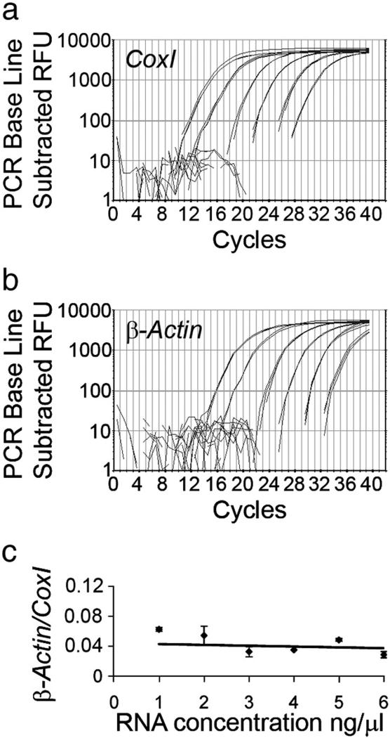
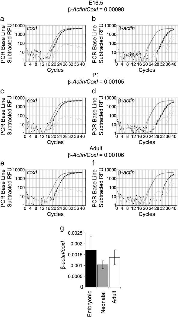
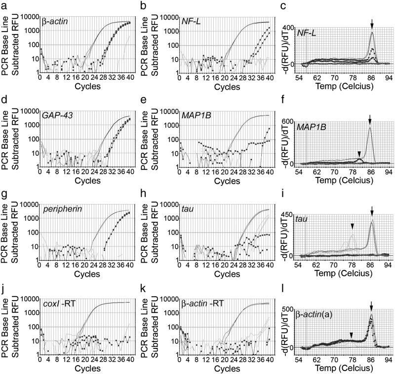
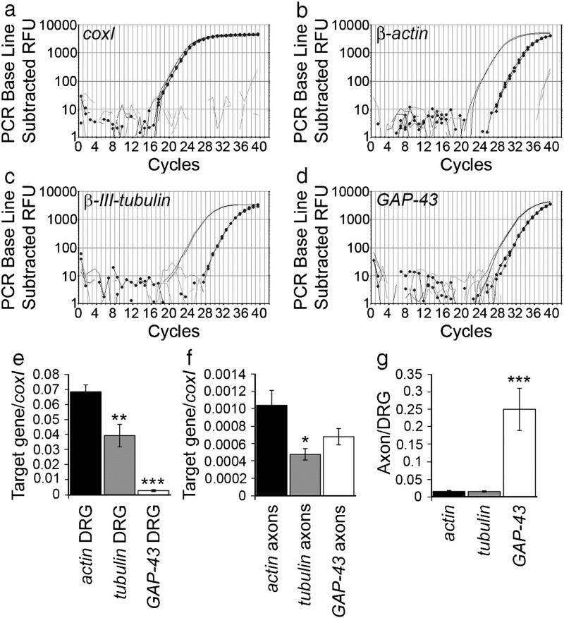
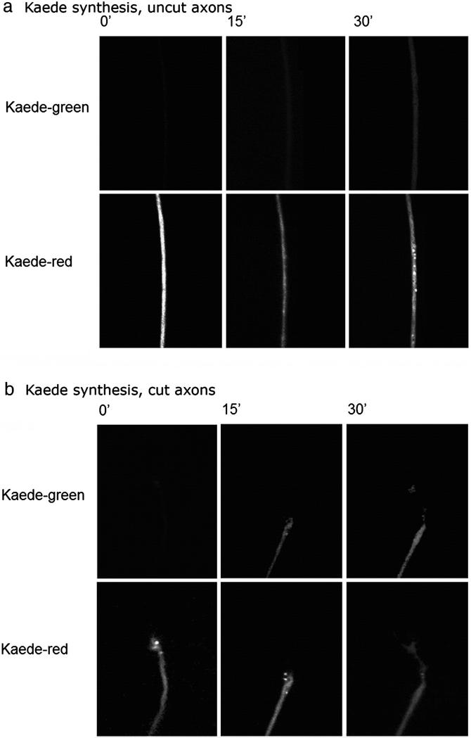
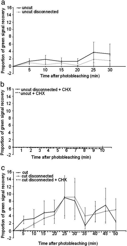
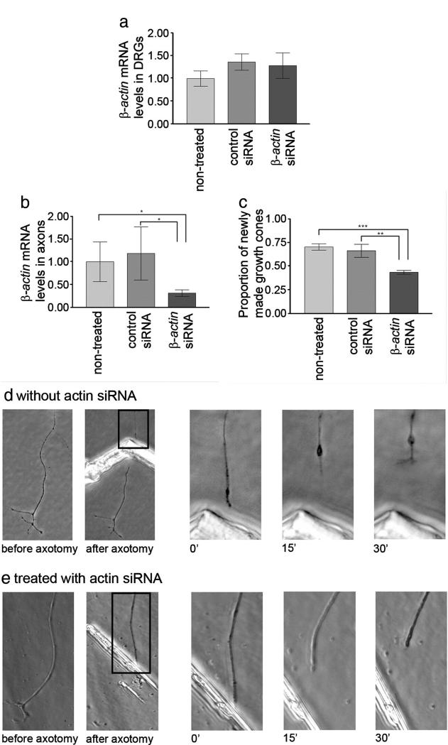
We have developed a compartmentalised culture model for the purification of axonal mRNA from embryonic, neonatal and adult rat dorsal root ganglia. This mRNA was used un-amplified for RT-qPCR. We assayed for the presence of axonal mRNAs encoding molecules known to be involved in axon growth and guidance. mRNAs for beta-actin, beta-tubulin, and several molecules involved in the control of actin dynamics and signalling during axon growth were found, but mRNAs for microtubule-associated proteins, integrins and cell surface adhesion molecules were absent. Quantification of beta-actin mRNA by means of qPCR showed that the transcript is present at the same level in embryonic, newborn and adult axons. Using the photoconvertible reporter Kaede we showed that there is local translation of beta-actin in axons, the rate being increased by axotomy. Knock down of beta-actin mRNA by RNAi inhibited the regeneration of new axon growth cones after in vitro axotomy, indicating that local translation of actin-related molecules is important for successful axon regeneration.


Figures

Fig. 1
Fig. 1
Schematic representation of current compartment culture models I–IV (Eng et al., 1999; Zheng et al., 2001; Wu et al., 2005; Taylor et al., 2006). Our newly developed “explant chamber” is shown in (V).
Fig. 2
Fig. 2
P1 DRGs cultured in the compartmentalised explant chamber. (a) Explants were plated in a row on top of the scratches and the silicon insert placed perpendicular to the scratches. (b) Solutions placed in one compartment remain in that compartment; india ink was placed in the central compartment, the experiment photographed 4 h later. (c) Axons extended from the explants, crossed the insert and grew into the outer compartment, using the scratches for guidance. Axons were harvested from the outer compartment after 7–9 days. (c) P1 DRG axons in culture at 5–7.5 mm from the insert. Numerous axons can be seen lining up with the scratches. No cells were observed in most cultures. Scale bar=0.1 mm.
Fig. 3
Fig. 3
Sensitivity of the RT-qPCR for the detection of β-actin and coxI. RT-qPCR curves on DRG total RNA dilution series from 100 ng to 1 pg (in the graphs from left to right). Both coxI (a) and β-actin (b) were reliably and reproducibly amplified even from 1 pg of total RNA. Efficiencies of the coxI and β-actin PCRs were 95 and 90%, respectively. (c) Quantification of β-actin levels relative to coxI resulted in similar values for all RNA concentrations. The data points did not significantly differ from the average 0.044 (trendline) (ANOVA, p>0.05).
Fig. 4
Fig. 4
Quantification of β-actin mRNA in embryonic, neonatal and adult rat axon-only preparations. Examples of qPCR graphs for E16.5 (a, b), P1 (c, d), and adult (e, f) axon preparations. The positive control is shown in plain black, the axon-only preparation in dotted black, and the water control in grey. (g) Quantification of β-actin (b, d, f) levels relative to coxI (a, c, e) showed no significant difference between the developmental stages (ANOVA p=0.35).
Fig. 5
Fig. 5
Examples of RT-qPCR curves in axon-only preparations with similar coxI levels for 3 positive target mRNAs and 3 target mRNAs that were not detectable or did not match the selection criteria. The positive control is shown in plain black, the axon-only preparation in dotted black, and the water control in grey. β-Actin (a), GAP-43 (d) and peripherin (g) mRNAs were reliably detected in axon-only preparations. NF-L (b) did not match inclusion criteria, as the curves were not reproducible and the Ct values were too high. MAP1B (e) was not detected, although the positive control shows that it is highly abundant in the DRG itself. Tau (h) was not detectable. No signal was detected in minus RT controls for CoxI (j) and β-actin (k). Melt curves (c, f, i, l) were performed in order to confirm that the axonal product peak matches with the positive control product peak (arrows). The water controls in the β-actin (l) and tau (i) PCRs contain primer dimers (arrowheads) only.
Fig. 6
Fig. 6
Quantification of β-actin, β-III-tubulin and GAP-43 mRNA in neonatal rat axon-only preparations. Examples of qPCR graphs for β-actin (b), β-III-tubulin (c) and GAP-43 (d). The positive control is shown in plain black, the axon-only preparation in dotted black, and the water control in grey. Quantification relative to coxI (a) showed that β-III-tubulin mRNA levels were about half the levels of β-actin, in both DRGs (**p<0.01) and axons (*p<0.05) (e, f). GAP-43 mRNA, in contrast, was about 25 times lower than actin in DRGs (***p<0.0001), but only 1.5 times lower than actin in axons (e, f). Axonal levels of actin and tubulin were about 1.5% and 1.3% respectively of the DRG levels, whereas, the levels of GAP-43 in axons was 25% of DRG levels (g).
Fig. 7
Fig. 7
Synthesis of Kaede driven by 3′UTR of rat β-actin in axons. At time 0 all the Keade is photobleached to red, then newly synthesised Kaeda appears in the green channel. In cut axons the newly synthesised Kaede is detected more rapidly and at a higher level.
Fig. 8
Fig. 8
Local synthesis of β-actin measured by recovery of photoconverted Kaede driven by 3′UTR of rat β-actin. (a) Kaede green signal recovery in uncut axons. Recovery of green signal was observed in uncut axons attached to, but also disconnected from the cell body, demonstrating that β-actin protein is produced by local axonal synthesis. No significant difference between attached or disconnected axons was observed. (b) Recovery of green signal in uncut axons was abolished by treatment with CHX (c) Axotomy increases the rate of local β-actin synthesis in regenerating axons attached to, but also disconnected from the cell body. Regression analysis showed that the rate of synthesis is greater in cut axons compared to the uncut axons shown in (a) (97.5% confidence), and in disconnected cut axons compared to disconnected uncut axons (99% confidence). Recovery of green signal in regenerating axons expressing Kaede-β-actin 3′ UTR was abolished by treatment with CHX.
Fig. 9
Fig. 9
Knock down of β-actin mRNA in axons reduces the ability of axons to regenerate after axotomy in vitro. β-actin siRNA treatment delivered to the axonal compartment did not reduce the β-actin mRNA levels (levels shown relative to non-treated samples) in the DRGs in the cell body compartment (a), but did reduce β-actin mRNA levels in the axons T-test *p<0.05 (b). (c) The number of regenerating axons is shown as a proportion of axomotised axons. Treatment of the axonal compartment with β-actin siRNA reduced the ability of axons to form a new growth cone after axotomy in vitro. Treatment with a control siRNA does not lead to significant decrease of regeneration ability. ANOVA, Bonferroni post-test **p<0.01 ***p<0.001, error bars are SEM. (d and e) show representative experiments in which axons that either had or had not been treated with β-actin siRNA were cut. In (d) the axon retracts with a retraction bulb, then generates a new growth cone within 30 min, while the treated axon in (e) retracts but does not form a new growth cone.

References

    1. Aronov S, Aranda G, Behar L, Ginzburg I. Axonal tau mRNA localization coincides with tau protein in living neuronal cells and depends on axonal targeting signal. J. Neurosci. 2001;21:6577–6587. - PMC - PubMed
    1. Bassell GJ, Zhang HL, Byrd AL, Femino AM, Singer RH, Taneja KL, Lifshitz LM, Herman IM, Kosik KS. Sorting of beta-actin mRNA and protein to neurites and growth cones in culture. J. Neurosci. 1998;18:251–265. - PMC - PubMed
    1. Burden-Gulley SM, Payne HR, Lemmon V. Growth cones are actively influenced by substrate-bound adhesion molecules. J. Neurosci. 1995;15:4370–4381. - PMC - PubMed
    1. Chierzi S, Ratto GM, Verma P, Fawcett JW. The ability of axons to regenerate their growth cones depends on axonal type and age, and is regulated by calcium, cAMP and ERK. Eur. J. Neurosci. 2005;21:2051–2062. - PubMed
    1. Chun JT, Gioio AE, Crispino M, Giuditta A, Kaplan BB. Differential compartmentalization of mRNAs in squid giant axon. J. Neurochem. 1996;67:1806–1812. - PubMed

Publication types

MeSH terms