. 2009 Jun;84(6):792-800.
doi: 10.1016/j.ajhg.2009.05.007.

Mutations in a BTB-Kelch protein, KLHL7, cause autosomal-dominant retinitis pigmentosa

Affiliations

Mutations in a BTB-Kelch protein, KLHL7, cause autosomal-dominant retinitis pigmentosa

James S Friedman et al. Am J Hum Genet. 2009 Jun.

Abstract

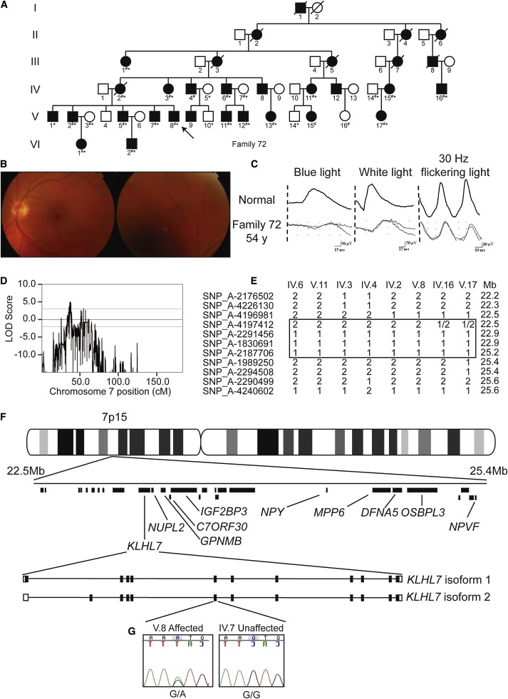
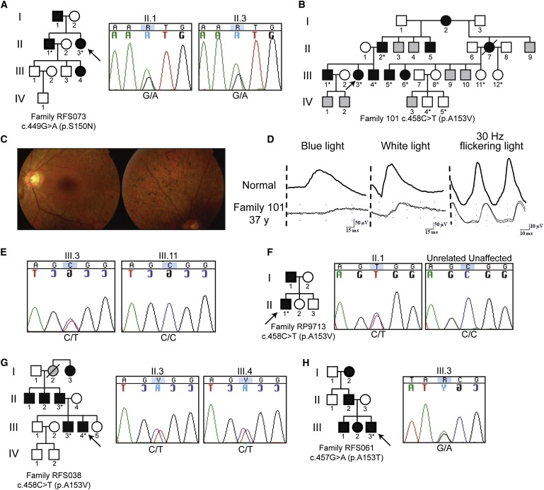
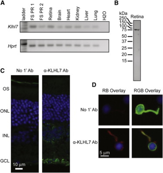
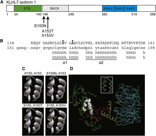
Retinitis pigmentosa (RP) refers to a genetically heterogeneous group of progressive neurodegenerative diseases that result in dysfunction and/or death of rod and cone photoreceptors in the retina. So far, 18 genes have been identified for autosomal-dominant (ad) RP. Here, we describe an adRP locus (RP42) at chromosome 7p15 through linkage analysis in a six-generation Scandinavian family and identify a disease-causing mutation, c.449G-->A (p.S150N), in exon 6 of the KLHL7 gene. Mutation screening of KLHL7 in 502 retinopathy probands has revealed three different missense mutations in six independent families. KLHL7 is widely expressed, including expression in rod photoreceptors, and encodes a 75 kDa protein of the BTB-Kelch subfamily within the BTB superfamily. BTB-Kelch proteins have been implicated in ubiquitination through Cullin E3 ligases. Notably, all three putative disease-causing KLHL7 mutations are within a conserved BACK domain; homology modeling suggests that mutant amino acid side chains can potentially fill the cleft between two helices, thereby affecting the ubiquitination complexes. Mutations in an identical region of another BTB-Kelch protein, gigaxonin, have previously been associated with giant axonal neuropathy. Our studies suggest an additional role of the ubiquitin-proteasome protein-degradation pathway in maintaining neuronal health and in disease.


Figures

Figure 1
Figure 1
Linkage Analysis and Genetic Screen of Family 72 (A) Pedigree of family 72. Individuals genotyped for linkage are noted by number symbols. Individuals sequenced in exon 6 of KLHL7 are noted with an asterisk. (B) Fundus photograph of individual V.8. Very mild degeneration is observed. (C) ERG traces of individual V.8 illustrate isolated rod responses to single flashes of blue light (left column), mixed responses to single flashes of white light (middle column), and isolated cone responses to 30 Hz flickering light (right column). (D) Linkage analysis of 23 individuals from family 72. A peak multipoint LOD score of 5.0 was obtained on chromosome 7p15. (E) Disease-chromosome haplotypes of individuals IV.6, V.11, IV.3, IV.4, IV.2, V.8, IV.16, and V.17. Recombinations between SNP_A-4196981 (rs4719697) and SNP_A-4197412 (rs3857716), and SNP_A-2187706 (rs7780038) and SNP_A-1989250 (rs2188993), respectively, denote the critical region. Mb denotes Megabase. (F) Physical map of the adRP critical region. Mb denotes Megabase. Genes completely screened for mutations are labeled. The intron-exon structure for KLHL7 isoforms 1 and 2 are shown. (G) Chromatogram of KLHL7 exon 6 sequences in individuals V.8 and IV.7 from family 72 are shown. Proband V.8 contains a heterozygous G-to-A transition, c.449G→A, leading to a predicted p.S150N amino acid change. A related unaffected individual, IV.7, has a homozygous G at the same residue.
Figure 2
Figure 2
Mutation Screen of KLHL7 Individuals in pedigrees sequenced in exon 6 of KLHL7 are denoted with an asterisk. Probands are marked with an arrow. Individuals of unknown phenotype are marked with gray. (A) Pedigree and chromatograms of a c.449G→A-containing proband in North American family RFS073. Sequences of II.1 and II.3 are shown. (B) Pedigree of Scandinavian adRP family 101. (C) Fundus photograph of individual III.3. Retinal degeneration and pigmentation are observed. (D) ERG analysis of individual III.3. Rod and cone photoreceptor responses were moderately reduced. (E) Chromatograms of individuals III.3 and III.11, showing the c.458C→T mutation and no change, respectively. (F) Pedigree of UK family RP9713. Chromatogram of individual II.1, with the c.458C→T change, and an unrelated unaffected individual. (G) Pedigree of North American adRP family RFS038. Chromatograms of individuals II.3, III.3, and III.4 reveal the c.458C→T mutation. (H) Pedigree of North American adRP family RFS061. Chromatogram of proband III.3, showing the c.457G→A mutation.
Figure 3
Figure 3
Expression Analysis and Protein Localization of KLHL7 (A) Klhl7 RT-PCR product was observed in cDNA from various tissues (as indicated) and GFP-tagged flow-sorted rod photoreceptors (FS PR1 and FS PR2). (B) Immunoblot analysis of KLHL7. A protein of approximately 75 kDa was observed in mouse retina with the use of an α-KLHL7 antibody. Predicted molecular mass of KLHL7 is ∼65 kDa. (C) Immunohistochemical characterization of KLHL7 protein. Mouse retina was incubated with secondary antibody only (left panel) or with α-KLHL7 (right panel). KLHL7 staining (green) is strong around the nuclei in the ganglion cell and the inner nuclear layer. (D) Immunocytochemistry of a mouse rod photoreceptor. GFP-tagged photoreceptors were dissociated from Nrl-GFP mouse retina and incubated with an α-KLHL7 antibody. Weak KLHL7 staining (red) is detected around the nuclei (DAPI blue) of photoreceptors.
Figure 4
Figure 4
KLHL7 Protein Structure and Analysis (A) Schematic of KLHL7 isoform 1. Mutations, reported here, are indicated. (B) Protein alignment of the KLHL7 BACK domain to Kbtbd4. Mutant residues S150 and A153 are shown in bold and marked with an asterisk. (C) Helical model of the KLHL7 BACK domain. S150 and A153 side chains are purple and green, respectively. Mutations 150N, 153T, and 153V are red, blue, and yellow, respectively. Each mutant side chain appears to occupy more space than the respective wild-type side chain in the cleft between two helices. (D) Superimposition of α1 and α2 helices of the KLHL7 BACK domain on the Skp1 complex. The α1 and α2 helices of the KLHL7 BACK domain are structurally similar (RMSD = 0.84Å) to the α5 and α6 helices of Skp1 (inset). Green indicates Skp1. White is used to show an overlay of the BACK-domain α1 and α2 to α5 and α6 helices of Skp1. Red indicates the F box. Yellow indicates the helical linker. Blue indicates the WD40 domain. PDB:1nex.

References

    1. Hartong D.T., Berson E.L., Dryja T.P. Retinitis pigmentosa. Lancet. 2006;368:1795–1809. - PubMed
    1. Daiger S.P., Bowne S.J., Sullivan L.S. Perspective on genes and mutations causing retinitis pigmentosa. Arch. Ophthalmol. 2007;125:151–158. - PMC - PubMed
    1. Sullivan L.S., Bowne S.J., Birch D.G., Hughbanks-Wheaton D., Heckenlively J.R., Lewis R.A., Garcia C.A., Ruiz R.S., Blanton S.H., Northrup H. Prevalence of disease-causing mutations in families with autosomal dominant retinitis pigmentosa: A screen of known genes in 200 families. Invest. Ophthalmol. Vis. Sci. 2006;47:3052–3064. - PMC - PubMed
    1. Sancho-Pelluz J., Arango-Gonzalez B., Kustermann S., Romero F.J., van Veen T., Zrenner E., Ekstrom P., Paquet-Durand F. Photoreceptor cell death mechanisms in inherited retinal degeneration. Mol. Neurobiol. 2008;38:253–269. - PubMed
    1. Ruschendorf F., Nurnberg P. ALOHOMORA: A tool for linkage analysis using 10K SNP array data. Bioinformatics. 2005;21:2123–2125. - PubMed

Publication types

MeSH terms