

Positron emission tomography imaging of (2R,3R)-5-[(18)F]fluoroethoxybenzovesamicol in rat and monkey brain: a radioligand for the vesicular acetylcholine transporter
- PMID: 19520289
- PMCID: PMC2696062
- DOI: 10.1016/j.nucmedbio.2009.02.007
Positron emission tomography imaging of (2R,3R)-5-[(18)F]fluoroethoxybenzovesamicol in rat and monkey brain: a radioligand for the vesicular acetylcholine transporter
Abstract
Introduction: The regional brain distribution of (2R,3R)-5-[(18)F]fluoroethoxy-benzovesamicol ((-)-[(18)F]FEOBV), a radioligand for the vesicular acetylcholine transporter (VAChT), was examined in vivo in mice, rats and rhesus monkeys.
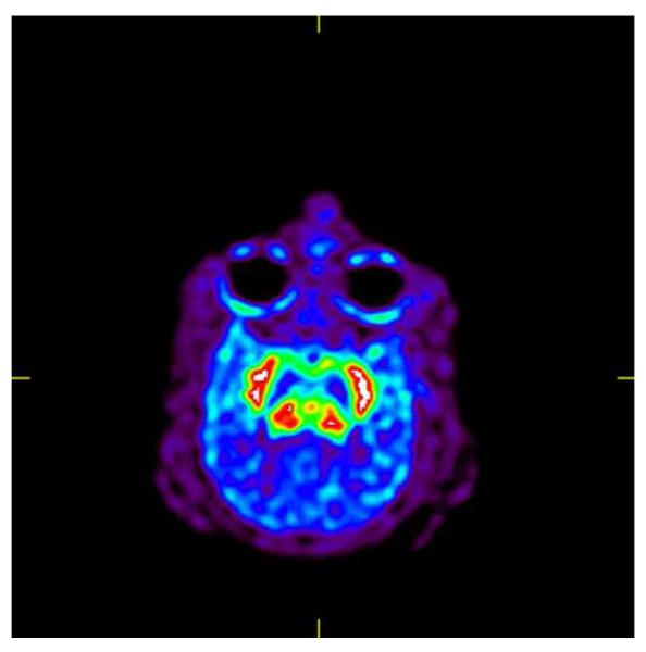
Methods: Regional brain distributions of (-)-[(18)F]FEOBV in mice were determined using ex vivo dissection. MicroPET imaging was used to determine the regional brain pharmacokinetics of the radioligand in rat and rhesus monkey brains.
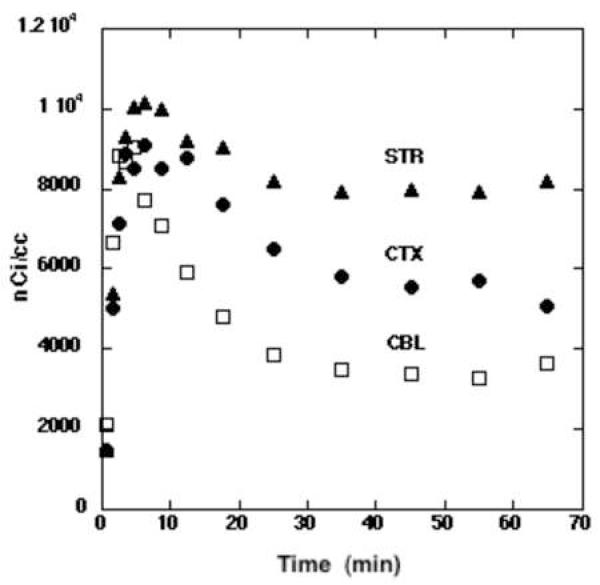
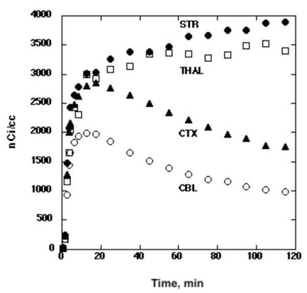
Results: In all three species, clear heterogeneous regional brain distributions were obtained, with the rank order of brain tissues (striatum>thalamus>cortex>cerebellum) consistent with the distribution of cholinergic nerve terminals containing the VAChT.
Conclusions: (-)-[(18)F]FEOBV remains a viable candidate for further development as an in vivo imaging agent for positron emission tomography (PET) studies of the VAChT in the human brain.
Figures
References
-
- Parsons SM, Prior C, Marshall IG. Acetylcholine transport, storage and release. Int Rev Neurobiol. 1993;35:279–390. - PubMed
-
- Frey KA, Wieland DM, Kilbourn MR. Imaging of monoaminergic and cholinergic vesicular transporters in the brain. In: Goldstein D, Eisenhofer G, McCarty R, editors. Catecholamines: Bridging Basic Science with Clinical Medicine. Academic Press; San Diego: 1997. pp. 269–272.
-
- Efange SM. In vivo imaging of the vesicular acetylcholine transporter and the vesicular monoamine transporter. Faseb J. 2000;14:2401–13. - PubMed
-
- Kuhl DE, Minoshima S, Fessler JA, Frey KA, Foster NL, Ficaro EP, Wieland DM, Koeppe RA. In vivo mapping of cholinergic terminals in normal aging, Alzheimer’s disease, and Parkinson’s disease. Ann Neurol. 1996;40:399–410. - PubMed
-
- Albin RL, Cross D, Cornblath WT, Wald JA, Wernette K, Frey KA, Minoshima S. Diminished striatal [123I]iodobenzovesamicol binding in idiopathic cervical dystonia. Ann Neurol. 2003;53:528–32. - PubMed
MeSH terms
Substances
Grants and funding
LinkOut - more resources
Full Text Sources
