

[(186)Re]Liposomal doxorubicin (Doxil): in vitro stability, pharmacokinetics, imaging and biodistribution in a head and neck squamous cell carcinoma xenograft model
- PMID: 19520292
- PMCID: PMC2696057
- DOI: 10.1016/j.nucmedbio.2009.02.004
[(186)Re]Liposomal doxorubicin (Doxil): in vitro stability, pharmacokinetics, imaging and biodistribution in a head and neck squamous cell carcinoma xenograft model
Abstract
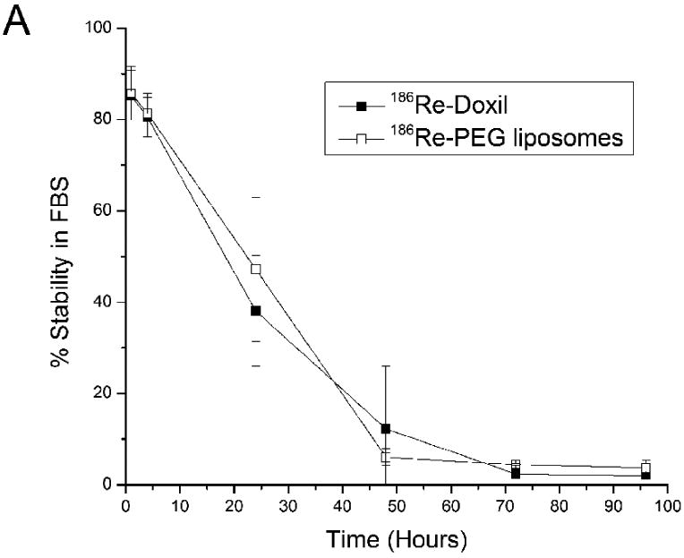
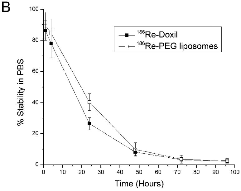
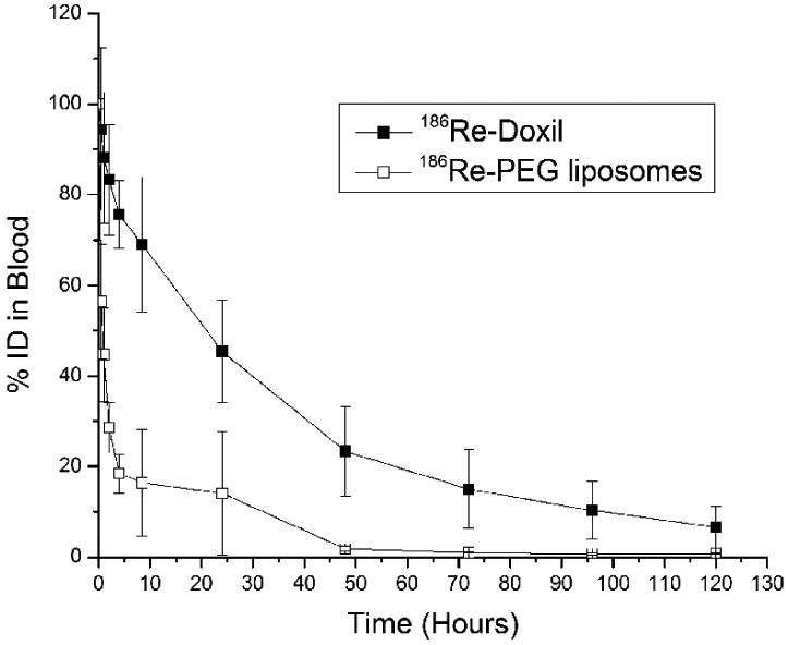
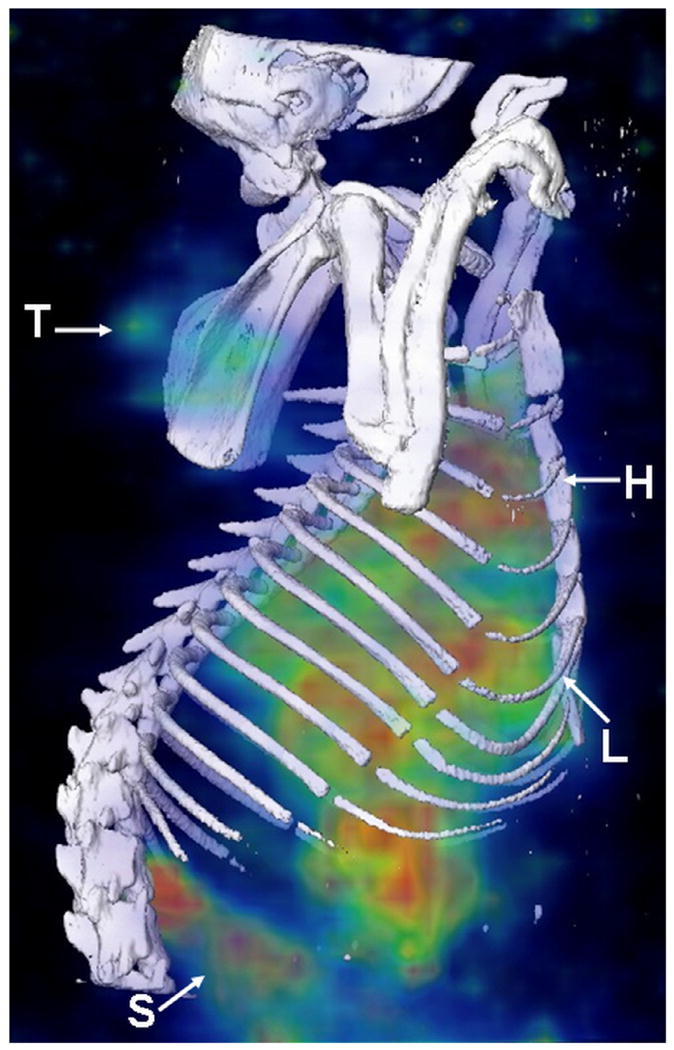
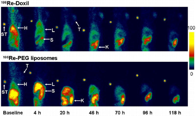
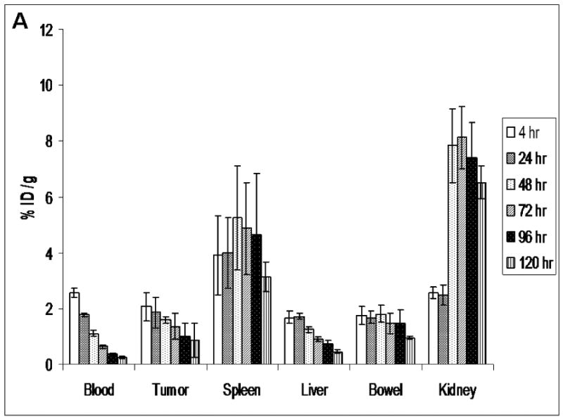
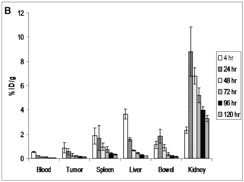
The purpose of this study was to determine the feasibility of radiolabeling liposomal doxorubicin (Doxil) for cancer chemoradionuclide therapy by directly loading the therapeutic radionuclide rhenium-186 ((186)Re) into the liposome interior. The pharmacokinetics, imaging and biodistribution of [(186)Re]Doxil (555 MBq/kg) and control [(186)Re]polyethylene glycol (PEG) liposomes (555 MBq/kg) were determined after intravenous administration in a head and neck cancer xenograft model in nude rats. [(186)Re]Doxil and [(186)Re]PEG liposomes were radiolabeled using [(186)Re]N,N-bis(2-mercaptoethyl)-N',N'-diethylethylenediamine. (186)Re labeling efficiency was 76.1+/-8.3% with Doxil. The in vitro serum stability of [(186)Re]Doxil at 37 degrees C was 38.06+/-12.13% at 24 h. Pharmacokinetic studies revealed that [(186)Re]Doxil had a two-phase blood clearance with half clearance times of 0.8 and 28.2 h. Images acquired over 120 h showed that [(186)Re]Doxil had slow blood clearance, low liver accumulation and increasing spleen accumulation. The biodistribution study at 120 h indicated that the percentage of injected dose (%ID) in the blood and tumor for [(186)Re]Doxil was 20-fold higher than that of [(186)Re]PEG liposomes. The %ID values in the kidney and liver were not significantly different between [(186)Re]Doxil and [(186)Re]PEG liposomes. These results suggest that the long circulation and prolonged bioavailability of [(186)Re]Doxil could potentially deliver high concentrations of both doxorubicin and (186)Re to tumor when encapsulated in the same liposome vehicle.
Figures
References
-
- Gabizon AA. Pegylated liposomal doxorubicin: metamorphosis of an old drug into a new form of chemotherapy. Cancer Invest. 2001;19:424–36. - PubMed
-
- Gabizon A, Shmeeda H, Barenholz Y. Pharmacokinetics of pegylated liposomal Doxorubicin: review of animal and human studies. Clin Pharmacokinet. 2003;42:419–36. - PubMed
-
- Gabizon AA, Barenholz Y, Bialer M. Prolongation of the circulation time of doxorubicin encapsulated in liposomes containing a polyethylene glycol-derivatized phospholipid: pharmacokinetic studies in rodents and dogs. Pharm Res. 1993;10:703–8. - PubMed
-
- Gabizon AA, Shmeeda H, Zalipsky S. Pros and cons of the liposome platform in cancer drug targeting. J Liposome Res. 2006;16:175–83. - PubMed
-
- Parkin DM, Bray F, Ferlay J, Pisani P. Estimating the world cancer burden: Globocan 2000. Int J Cancer. 2001;94:153–6. - PubMed
Publication types
MeSH terms
Substances
Grants and funding
LinkOut - more resources
Full Text Sources
Other Literature Sources
Medical
Miscellaneous
