. 2009 Sep;132(3):362-70.
doi: 10.1016/j.clim.2009.05.011. Epub 2009 Jun 10.

T cell CD40LG gene expression and the production of IgG by autologous B cells in systemic lupus erythematosus

Affiliations

T cell CD40LG gene expression and the production of IgG by autologous B cells in systemic lupus erythematosus

Ying Zhou et al. Clin Immunol. 2009 Sep.

Abstract

CD40 ligand (CD40LG), encoded on the X chromosome, has been reported to be overexpressed on lupus T cells. Herein, we investigated the effect of DNA demethylation on T cell CD40LG expression and the production of IgG by autologous B cells in lupus. We found normal human T cells transfected with CD40LG induced autologous B cell activation and plasma cell differentiation. Both female lupus CD4+ T cells and demethylating agents treated CD4+ T cells overexpressed CD40LG mRNA. Further, lupus T cells from both genders or demethylated CD4+ T cells from healthy women overstimulated autologous B cells, and this could be reversed with anti-CD40LG Ab in only females. We demonstrated that female lupus CD4+ T cells and demethylated CD4+ T cells express high level of CD40LG and overstimulate B cells to produce IgG. This is due to DNA demethylation and thereby reactivation of the inactive X chromosome in female.


Figures

Figure 1
Figure 1
(A) Increased surface expression of CD40LG on human T cells transfected with pEGFP-C1/CD40LG compared to empty vector transfected controls. (B) Increased percentage of CD25 expression (P=0.0002, n=7), (C) CD69 expression (P=0.0041, n=6), and (D) CD138 expression (P=0.0096, n=7) on CD19+ gated cells that were cocultured with CD40LG overexpressing T cells as compared to T cells transfected with empty vector. Representative histograms are shown on the right.
Figure 2
Figure 2
CD40LG mRNA expression on CD4+ and CD8+ T cells. CD40LG transcripts relative β-actin was measured using realtime RT-PCR. Results represent the mean±SEM of the indicated numbers of subjects. (A) in women (n=6), T cells from lupus patients expressed significantly higher amount of CD40LG mRNA than normal controls (n=6), *=P<0.05 versus normal controls (CD4+, P=0.020, CD8+, P=0.045). As in men, CD40LG expression on CD4+ and CD8+ T cells was not significantly different between patients with lupus (n=9) and normal controls (n=5) (P>0.05). (B) in DNA methylation inhibitors treated or untreated T cells. Treated CD4+ T cells from women expressed significantly increased CD40LG mRNA compared with untreated CD4+ T cells from women (n=3), *=P<0.05 versus untreated CD4+ T cells (5-AzaC, P=0.002; procainamide, P=0.047; hydralazine, P=0.039 and PD98059, P=0.048). The difference was also significant between values of treated CD4+ T cells from women and treated CD4+ T cells from men(n = 3), ▲=P<0.05 versus CD4+ T cells from men(5-AzaC, P=0.009; hydralazine, P=0.046; procainamide, P=0.032 and PD98059 P=0.025).
Figure 3
Figure 3
IgG production induced by T–B cells culture in vitro. Controls included B cells cultured alone, B cells plus lipopolysaccharide (LPS), and B cells plus LPS and anti-CD40LG. (A) women group: IgG production of B cell cultured with autologous CD4+ or CD8+ T cell from female lupus was significantly higher in lupus patients. Lupus patients (n=6) versus normal controls (n=6) (P=0.000, P=0.000, respectively); * = P<0.05 versus normal controls. CD4+ T plus B cells with versus without anti-CD40LG: P=0.034; CD8+ T plus B cells with versus without mAb: P=0.047; **=P<0.05 versus cultures without pretreatment of T cells with anti-CD40LG. CD4+ T plus B cells versus B cells alone: P=0.011, CD8+ T plus B cells versus B cells alone: P=0.012; ▲=P<0.05 versus B cells cultured alone. (B) men group: IgG level of B cell cultured with autologous CD4+ or CD8+ T cell from female lupus was also significantly higher in lupus patients (n=9) than that in normal controls (n=5) (P=0.000, P=0.000, respectively) normal controls, *=P<0.05 versus normal controls. CD4+ T plus B cells plus with versus without anti-CD40LG: P=0.238; CD8+ T plus B cells with versus without anti-CD40LG: P=0.607. CD4+ T plus B cells versus B cells alone: P=0.000, CD8+ T plus B cells versus B cells: P=0.000.
Figure 4
Figure 4
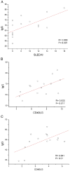
(A) Correlation between the IgG production and disease activity as measured by SLE disease index score analyses for all patients (R=0.668, P=0.007). (B) IgG secreted by coculturing CD4+ T cells and autologous B cells in vitro is plotted against the expression of CD40LG mRNA on CD4+ T cells from 15 lupus patients. Where indicated, IgG was measured by ELISA as described in the text. CD40LG mRNA was detected by real-time-PCR. (R=0.605, P=0.017). (C) Correlation of CD40LG mRNA on 5-AzaC, procainamide, hydralazine and PD98059 treated[Float1] CD4+ T cells from women and IgG secreted by coculturing 5-AzaC, procainamide, hydralazine and PD98059 treated CD4+ T cells and autologous B cells (R=0.641, P=0.01).
Figure 5
Figure 5
IgG synthesis induced by B cell cocultured with autologous demethylated T cell in vitro. CD4+ and CD8+ T cells from normal controls were pretreated with DNA methylation inhibitors and cultured with autologous B cells, controls included B cells cultured alone, B cells plus LPS, and B cells plus LPS and anti-CD40LG. (A) IgG was significantly promoted by CD4+ T cells pretreated with 5-AzaC (P=0.02), procainamide (P=0.026), hydralazine (P=0.013) and PD98059 (P=0.008) compared with untreated CD4+ T cells, ▲ =P<0.05 versus untreated CD4+ T cells. Anti-CD40LG inhibited IgG synthesis induced by CD4+ T treated with 5-AzaC (P=0.038), procainamide (P=0.042), hydralazine (P=0.024) and PD98059 (P=0.003), *=P<0.05 versus cultures with and without pretreatment of T cells with anti-CD40LG. Demethylated CD8+ T cells did not promote IgG production to a statistically significant level compared with untreated CD8+ T cell (P>0.05). Anti-CD40LG mAb inhibition of IgG was slightly on CD8+ T cells. (P=0.775) (B) drug-treated CD4+ or CD8+ T cells from men cultured with autologous B cells with or without anti-CD40LG mAb did not significantly affect IgG production. (C) CD4+ T cells from women pretreated with 5-AzaC (P=0.025), procainamide (P=0.041), hydralazine (P=0.022) and PD98059 (P=0.02) promoted IgG production compared with CD4+ T cells from men. * = P<0.05 versus IgG production of drug-treated CD4+ T cocultured with B cell form men.

References

    1. Sawalha AH, Harley JB. Antinuclear autoantibodies in systemic lupus erythematosus. Curr Opin Rheumatol. 2004;16:534–540. - PubMed
    1. Soto ME, Vallejo M, Guillen F, Simon JA, Arena E, Reyes PA. Gender impact in systemic lupus erythematosus. Clin Exp Rheumatol. 2004;22:713–721. - PubMed
    1. Lockshin MD. Sex ratio and rheumatic disease: excerpts from an Institute of Medicine report. Lupus. 2002;11:662–666. - PubMed
    1. Vogel LA, Noelle RJ. CD40 and its crucial role as a member of the TNFR family. Semin Immunol. 1998;10:435–442. - PubMed
    1. Lu LF, Cook WJ, Lin LL, Noelle RJ. CD40 signaling through a newly identified tumor necrosis factor receptor-associated factor 2 (TRAF2) binding site. J Biol Chem. 2003;278:45414–45418. - PubMed

Publication types

MeSH terms