. 2009 Aug 7;284(32):21317-26.
doi: 10.1074/jbc.M109.015016. Epub 2009 Jun 11.

The Thermoplasma acidophilum LplA-LplB complex defines a new class of bipartite lipoate-protein ligases

Affiliations

The Thermoplasma acidophilum LplA-LplB complex defines a new class of bipartite lipoate-protein ligases

Quin H Christensen et al. J Biol Chem. .

Abstract

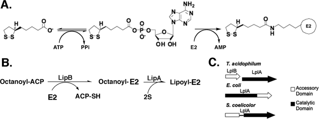
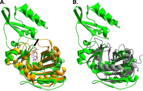
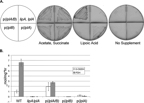
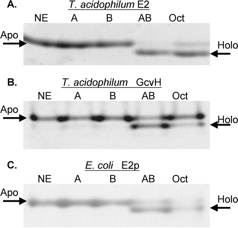
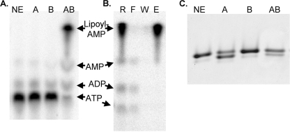
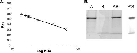
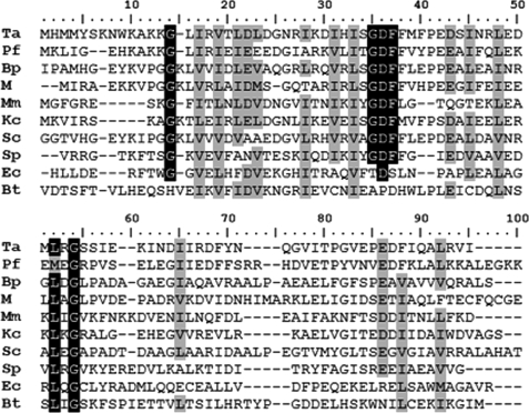
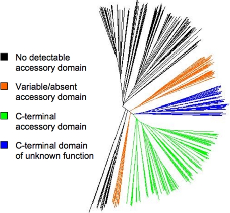
Lipoic acid is a covalently bound cofactor found throughout the domains of life that is required for aerobic metabolism of 2-oxoacids and for C(1) metabolism. Utilization of exogenous lipoate is catalyzed by a ligation reaction that proceeds via a lipoyl-adenylate intermediate to attach the cofactor to the epsilon-amino group of a conserved lysine residue of protein lipoyl domains. The lipoyl ligases of demonstrated function have a large N-terminal catalytic domain and a small C-terminal accessory domain. Half of the members of the LplA family detected in silico have only the large catalytic domain. Two x-ray structures of the Thermoplasma acidophilum LplA structure have been reported, although the protein was reported to lack ligase activity. McManus et al. (McManus, E., Luisi, B. F., and Perham, R. N. (2006) J. Mol. Biol. 356, 625-637) hypothesized that the product of an adjacent gene was also required for ligase activity. We have shown this to be the case and have named the second protein, LplB. We found that complementation of Escherichia coli strains lacking lipoate ligase with T. acidophilum LplA was possible only when LplB was also present. LplA had no detectable ligase activity in vitro in the absence of LplB. Moreover LplA and LplB were shown to interact and were purified as a heterodimer. LplB was required for lipoyl-adenylate formation but was not required for transfer of the lipoyl moiety of lipoyl-adenylate to acceptor proteins. Surveys of sequenced genomes show that most lipoyl ligases of the kingdom Archaea are heterodimeric. We propose that the presence of an accessory domain provides a diagnostic to distinguish lipoyl ligase homologues from other members of the lipoate/biotin attachment enzyme family.


Figures

FIGURE 1.
FIGURE 1.
Lipoic acid metabolism in E. coli. Panel A, the lipoyl ligase (LplA) reaction that proceeds through the lipoyl-adenylate intermediate. In E. coli LplA acts to scavenge lipoic acid from the growth medium. Panel B, schematic of lipoic acid synthesis in E. coli. LipB transfers an octanoyl moiety from the fatty acid biosynthetic intermediate, octanoyl-acyl carrier protein, to the LD domain of a lipoate-accepting protein (in this case the E2 subunit of a 2-oxoacid dehydrogenase). The octanoylated LD domain is the substrate of LipA, an S-adenosylmethionine radical enzyme that replaces one hydrogen atom on each of octanoate carbons 6 and 8 with sulfur atoms. Panel C, the differing arrangements of genes and domains found in lipoate ligases in T. acidophilum, E. coli, and Streptomyces coelicolor. Only a single nucleotide lies between the T. acidophilum LplB and LplA coding sequences.
FIGURE 2.
FIGURE 2.
Structural alignments of LplA and LipB structures. Previously published crystal structures were aligned using DeepView (37). Panel A, E. coli LplA (Protein Data Bank code 1X2H in green) aligned with T. acidophilum LplA (Protein Data Bank code 2ART in orange). The lipoyl-adenylate intermediate bound to T. acidophilum LplA is shown in purple. The adenylate binding loop is indicated with an arrow. Panel B, M. tuberculosis LipB (Protein Data Bank code 1W66 in gray) is aligned with the E. coli LplA structure of panel A. The purple line denotes the covalent decanoate adduct present in the M. tuberculosis LipB structure. The substrate binding pocket is conserved among members of the protein family. The accessory domain is not part of the binding pocket and appears to play an indirect role in catalysis.
FIGURE 3.
FIGURE 3.
Complementation of an E. coli lipoyl ligase null mutant strain by T. acidophilum lplA and lplB. The lipA lplA strain, TM131, was transformed with pBAD322G-derived plasmids, p(lplA), p(lplB), and p(lplA plus B), expressing the T. acidophilum genes (as indicated) from an arabinose-inducible promoter. The wild type (WT) and lipA lplA both with an empty vector are the control strains. The complementation assays were performed on M9 minimal agar containing 0.2% arabinose and 0.1% vitamin assay casamino acids. Where indicated, 5 mm acetate plus 5 mm succinate or 5 μg/ml lipoic acid was added. To prevent carryover of lipoic acid, before testing the strains were grown on the same medium containing 5 mm acetate, 5 mm succinate, and 24 mg/liter gentamicin. Panel A, ability of expression of LplA and LplB alone or together to restore growth of E. coli strain TM131 (lplA lipA) when supplemented with lipoic acid. Representative plates of three replicate experiments are shown. Panel B, activation of the pyruvate dehydrogenase (PDH) and 2-oxoglutarate dehydrogenase (OGDH) complexes upon expression of LplA and LplB alone or together was assayed using an acetylated NAD analogue as a substrate for the overall reaction of the dehydrogenase complex. Results are reported as μmol of NAD analogue reduced/mg of cell extract h−1. The error bars denote twice the S.D. from at least four assays.
FIGURE 4.
FIGURE 4.
Ligase activity of LplA and LplB with two acceptor LDs measured by the gel shift assay. The modified lipoyl domain loses a charge upon modification of the target lysine residue resulting in more rapid migration on native gels than the unmodified domain. Representative gels of three independent experiments are shown. Lane designations given in parentheses below are the same in all three panels. Lane 1, no enzyme (NE); lane 2, LplA (A); lane 3, LplB (B); lane 4, LplA plus LplB (AB); lane 5, LplA plus LplB with octanoate in place of lipoate (Oct). Reaction components are listed under “Experimental Procedures.” Panel A, modification of the T. acidophilum branched chain dehydrogenase E2 lipoyl domain by lipoate or octanoate attachment. Panel B, modification of the putative T. acidophilum GcvH. Panel C, modification of the E. coli hybrid E2p domain. Note that efficient lipoylation of the E. coli domain required a 16-fold longer incubation time than the T. acidophilum domains.
FIGURE 5.
FIGURE 5.
The role of LplB in synthesis of lipoyl-adenylate and transfer of the lipoyl moiety. Panel A, synthesis of 32P-labeled lipoyl-adenylate from [α-32P]ATP was analyzed by cellulose thin layer chromatography and visualized by autoradiography. Lane 1, no enzyme (NE); lane 2, LplA (A); lane 3, LplB (B); lane 4, LplA plus LplB (AB). Panel B, demonstration of enzyme-bound lipoyl-AMP with a centrifugal filter device. Lane 1, reaction (R); lane 2, flow-through (F); lane 3, final wash (W); lane 4, retained enzyme fraction (E). Panel C, transfer of lipoate from synthetic lipoyl-adenylate to LD assayed by gel shift. Lanes 1–4 are labeled as in panel A.
FIGURE 6.
FIGURE 6.
Characterization of the LplA-LplB complex. Panel A, size exclusion chromatography of the purified proteins. The calibration curve was prepared using (in order of ascending molecular mass) ribonuclease A, chymotrypsinogen, ovalbumin, and bovine serum albumin as standards, shown as “×” symbols. Hexahistidine-tagged LplA is shown as a circle and was estimated to have a mass of 16.6 ± 0.5 kDa, whereas mass spectrometry gave a mass of 30.8 kDa. Hexahistidine-tagged LplB eluted after the linear range of the column (<10 kDa), although it had a mass of 11.2 kDa as determined by mass spectrometry. The lipoyl ligase (LplA-LplB) complex is shown as a square and elutes soon after LplA and has an estimated mass of 18.2 ± 0.4 kDa and a mass calculated from the individual mass spectra of 41.1 kDa. The estimated sizes are the average of four runs with independent protein samples. The S.D. is also reported. Panel B, demonstration of the lipoyl ligase complex by Ni2+ affinity chromatography. The elution products were subjected to SDS-PAGE on a 4–20% gradient Tris-glycine gel and visualized with Coomassie Blue R-250. LplB copurified with hexahistidine-tagged LplA (lane AB). Hexahistidine-tagged LplA and LplB were also purified individually as references (lanes A and B, respectively). The rightmost lane is a phosphorimaging scan of l-[35S]methionine-labeled lipoyl ligase complex after SDS-PAGE on a 4–20% gradient Tris-glycine gel.
FIGURE 7.
FIGURE 7.
ClustalW alignment of representative LplB homologues. The first six sequences are representative single domain LplB homologues. Black shading denotes identical residues whereas gray shading denotes residues of similar properties. The putative S. coelicolor (Sc) ligase has an N-terminal LplB domain. E. coli LplA (Ec) is the canonical C-terminal domain ligase. The putative S. pneumoniae (Sp) LplA also has a C-terminal domain. The Bos taurus (Bt) bLT has a C-terminal domain of unknown function. Conserved Gly residues correspond to flexible loops in the E. coli LplA structure (10). Most conserved residues are predicted to be in the interior of the protein and probably serve structural roles. Close relatives to LplB, including single domain proteins, contain a GDFF motif. The aspartate residue of this motif (Asp-41 in LplB) is well conserved among LplB homologues. The Genbank accession numbers of the aligned sequences (all previously published) are (from top to bottom) NP_393989, NP_579363.1, NP_880065, YP_687213.1, ZP_02512737, ACB07333.1, CAA18910, NP_345629, AAC77339, and BAA24354. Ta, T. acidophilum; Pf, Pyrococcus furiosus; Bp, Bordetella pertussis; M, methanogen, uncultured; Mm, Mycoplasma mycoides; Kc, Korarchaeum cryptofilum.
FIGURE 8.
FIGURE 8.
The lipoyl ligase subtree of the BPL_LplA_LipB Pfam family (PF03099) displayed using Dendroscope (38). Domain architecture is annotated by color. Black clade proteins contain a catalytic domain but no detectable LplB homologue in the genome. The green clade proteins have a C-terminal accessory domain. Blue clade proteins are found only in mammalian genomes and have a C-terminal domain of unknown function that has only background sequence similarity with the green clade C-terminal accessory domains (Fig. 7). The orange clade proteins have a variable domain architecture including N-terminal accessory domains and independently coded accessory domains. The deeply branching and multiclade presence of LplAs with independent LplBs suggests that this architecture is an evolutionary relic. The tree root, determined with biotin ligases and octanoyltransferases as outgroups, is within the black clades. This suggests that the catalytic domain was originally independent of any other domains in the common ancestral protein.

References

    1. Perham R. N. (2000) Annu. Rev. Biochem. 69, 961–1004 - PubMed
    1. Engel N., van den Daele K., Kolukisaoglu U., Morgenthal K., Weckwerth W., Pärnik T., Keerberg O., Bauwe H. (2007) Plant Physiol. 144, 1328–1335 - PMC - PubMed
    1. Reed L. J., Leach F. R., Koike M. (1958) J. Biol. Chem. 232, 123–142 - PubMed
    1. Koike M., Reed L. J. (1960) J. Biol. Chem. 235, 1931–1938 - PubMed
    1. Green D. E., Morris T. W., Green J., Cronan J. E., Jr., Guest J. R. (1995) Biochem. J. 309, 853–862 - PMC - PubMed

Publication types

LinkOut - more resources