. 2010 Apr;42(4):432-41.
doi: 10.1165/rcmb.2009-0002OC. Epub 2009 Jun 11.

Phosphatidylinositol-3-kinase/akt regulates bleomycin-induced fibroblast proliferation and collagen production

Affiliations

Phosphatidylinositol-3-kinase/akt regulates bleomycin-induced fibroblast proliferation and collagen production

Yongju Lu et al. Am J Respir Cell Mol Biol. 2010 Apr.

Abstract

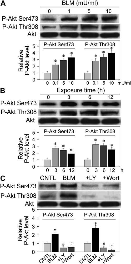
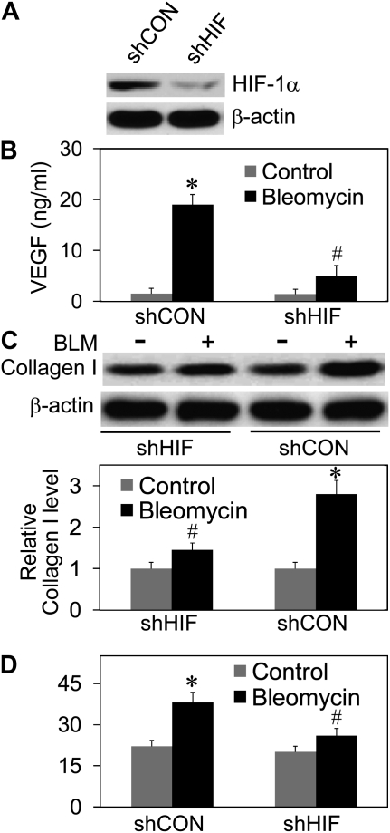
Abnormal repair and dysregulated angiogenesis have been implicated in the pathogenesis of pulmonary fibrosis, but the underlying mechanisms of regulation are not well understood. The present study investigated the role of phosphatidylinositol-3-kinase (PI3K)/Akt in fibrogenesis of human lung fibroblasts and its regulation by reactive oxygen species (ROS). Exposure of lung fibroblasts to bleomycin, a known inducer of fibrosis, resulted in rapid activation of PI3K/Akt and a parallel increase in fibroblast proliferation and collagen production, characteristics of lung fibrosis. Bleomycin had no significant effect on total Akt protein expression but induced phosphorylation of the protein at threonine 308 and serine 473 positions. Inhibition of this phosphorylation by PI3K inhibitors or by dominant-negative Akt (T308A/S473A) expression abrogated the effects of bleomycin on fibroblast proliferation and collagen production, suggesting the role of PI3K/Akt in the fibrogenic process. Activation of PI3K/Akt by bleomycin also led to transcriptional activation and protein expression of hypoxia-inducible factor-1alpha (HIF-1alpha) and vascular endothelial growth factor, which contributed to the fibroproliferative and collagen-inducing effects of bleomycin. The fibrogenic effects of bleomycin were dependent on ROS generation, particularly superoxide anion and hydrogen peroxide, which were induced by bleomycin. Inhibition of ROS generation by antioxidant enzymes, catalase and superoxide dismutase mimetic MnTBAP, abrogated the fibrogenic effects of bleomycin as well as its induction of PI3K/Akt and HIF-1alpha activation. Together, our results indicate a novel role of PI3K/Akt in fibrogenesis of human lung fibroblasts and its regulation by ROS, which could be exploited for the treatment of pulmonary fibrosis and related disorders.


Figures

Figure 1.
Figure 1.
Induction of fibroblast proliferation and collagen production by bleomycin. (A) Subconfluent cultures of human lung fibroblast CRL-1490 cells were exposed to various concentrations of bleomycin (0–25 mU/ml) and analyzed for cell growth after 24 hours. (B) Cells were similarly exposed to bleomycin for 24 hours and analyzed for apoptosis by enzyme-linked immunosorbent assay (ELISA)-based DNA fragmentation assay. (C) Western blot analysis of collagen protein expression in response to bleomycin treatment. Cells were treated with the indicated concentrations of bleomycin for 12 hours, after which they were washed with ice-cold phosphate-buffered saline (PBS) and extracted with SDS sample buffer. The cell extracts were separated on polyacrylamide-SDS gels, transferred, and probed with antibodies specific for human collagen type I and III. Blots were reprobed with β-actin antibody to confirm equal loading of the samples. The immunoblot signals were quantified by densitometry. Mean densitometry data from independent experiments (one of which is shown here) were normalized to the result obtained in cells in the absence of bleomycin (control). (D) Sircol assay of soluble collagen content in the supernatants of bleomycin-treated cells from above. Values are mean ± SD (n = 4). *P < 0.05 versus nontreated control.
Figure 2.
Figure 2.
Induction of Akt phosphorylation by bleomycin and its inhibition by phosphatidylinositol-3-kinase (PI3K) inhibitors. (A) Dose effect of bleomycin on Akt phosphorylation. CRL-1490 cells were treated with various concentrations of bleomycin for 6 hours, after which they were washed with PBS and extracted with SDS sample buffer. The cell extracts were separated on 10% polyacrylamide-SDS gels, transferred, and probed with antibodies against phospho-Akt (Ser473 and Thr308) and Akt. (B) Time-dependent effect of bleomycin on Akt phosphorylation. Cells were treated with bleomycin (10 mU/ml) for various times and analyzed for phospho-specific and total Akt by immunoblotting. (C) Cells were either left untreated or pretreated with LY294002 (10 μM) or wortmannin (10 μM) for 1 hour, followed by bleomycin treatment (10 mU/ml) for 6 hours. Cell lysates were prepared and analyzed for phospho-Akt and total Akt. The immunoblot signals were quantified by densitometry. Values are mean ± SD (n = 3). *P < 0.05 versus nontreated control, #P < 0.05 versus bleomycin-treated control.
Figure 3.
Figure 3.
Effects of PI3K and mitogen-activated protein kinase/ERK kinase (MEK) inhibitors on bleomycin-induced collagen production and fibroblast cell growth. (A) CRL-1490 cells were either left untreated or pretreated for 1 hour with the indicated concentrations of LY294002, wortmannin, or PD98059, followed by bleomycin treatment (10 mU/ml) for 12 hours. Collagen protein expression was determined by Western blotting. The density of collagen bands was quantified and normalized against the nontreated control band. Plots show treatment effect on collagen type I expression. Similar results were observed with collagen type III. (B) Cell supernatants from above were analyzed for soluble collagen content by Sircol assay. (C) Cells were treated with the test agents as described above and assayed for cell growth after 24 hours. (D) Cells were stably transfected with Akt mutant (SRα-Akt T308A/S473A) plasmid or control plasmid as described in Materials and Methods. Transfected cells were treated with bleomycin (10 mU/ml) for 12 hours and collagen expression, cell proliferation, and Akt phosphorylation were determined. Values are mean ± SD (n = 3). *P < 0.05 versus nontreated control, #P < 0.05 versus bleomycin-treated control.
Figure 4.
Figure 4.
Effects of bleomycin and PI3K inhibitors on vascular endothelial growth factor (VEGF) and hypoxia-inducible factor-1α (HIF-1α) expression. (A) CRL-1490 cells were treated with the indicated concentrations of bleomycin and at various times after the treatment, cell supernatants were collected and analyzed for VEGF protein by ELISA. (B) Cells were treated with bleomycin (10 mU/ml) in the presence or absence of LY294002 (10 μM). At various times after the treatment, VEGF protein levels were determined as described. (C) Cells were transfected with 1 μg of either the VEGF reporter (VEGF-luc) or control plasmid and 10 ng of pRL-tk normalizing luciferase plasmid. Transfected cells were treated with bleomycin (10 mU/ml) in the presence or absence of LY294002 (10 μM), and analyzed for luciferase activity after 12 hours. (D) RT-PCR was done to quantify the expression of the indicated transcripts after treatment of the cells with various concentrations of bleomycin (0–25 mU/ml) for 12 hours. (E) Immunoblot analysis of HIF-1α protein expression after treatment of the cells with the indicated concentrations of bleomycin for 12 hours. Cell extracts were prepared, separated on 10% polyacrylamide-SDS gels, transferred, and probed with HIF-1α antibody. (F) Cells were either left untreated or pretreated with LY294002 (10 μM) or wortmannin (10 μM) for 1 hour, followed by bleomycin treatment (10 mU/ml) for 12 hours. Cell lysates were prepared and analyzed for HIF-1α. Blots were reprobed with β-actin antibody to confirm equal loading of the samples. The immunoblot signals were quantified by densitometry. Values are mean ± SD (n = 3). *P < 0.05 versus nontreated control, #P < 0.05 versus bleomycin-treated control.
Figure 5.
Figure 5.
Effects of HIF-1α knockdown on bleomycin-induced VEGF secretion, fibroblast proliferation, and collagen expression. CRL-1490 cells were transfected with HIF-1α shRNA (shHIF) viral particles or control shRNA (shCON) particles as described in Materials and Methods. (A) Thirty-six hours after the transfection, cells were analyzed for HIF-1α expression by Western blotting. (B and C) Transfected cells were treated with bleomycin (10 mU/ml) for 12 hours and analyzed for VEGF secretion by ELISA and collagen expression by Western blotting. (D) Cell growth determined at 24 hours after bleomycin treatment (10 mU/ml). Values are mean ± SD (n = 3). *P < 0.05 versus nontreated control, #P < 0.05 versus shCON-treated control.
Figure 6.
Figure 6.
Effects of VEGF on collagen expression and fibroblast proliferation. (A) CRL-1490 cells were treated with recombinant VEGF (10 and 25 ng/ml) for 12 hours and analyzed for collagen expression by Western blotting. Bleomycin (10 mU/ml) was used as a positive control. Densitometry was performed to determine the relative levels of collagen expression after reprobing with β-actin antibody. (B) Cells were similarly treated with VEGF and bleomycin, and analyzed for cell growth after 24 hours. (C and D) Cells were treated with bleomycin (10 mU/ml) in the presence of neutralizing anti-VEGF or control antibody (100 ng/ml), and collagen expression and cell growth were determined after 12 hours and 24 hours, respectively. Values are mean ± SD (n = 4). *P < 0.05 versus nontreated control, #P < 0.05 less than control antibody treatment with bleomycin but greater than non–bleomycin-treated control.
Figure 7.
Figure 7.
Induction of reactive oxygen species (ROS) generation by bleomycin and its effects on Akt, HIF-1α, and VEGF expression. (A) CRL-1490 cells were treated with various concentrations of bleomycin (0–10 mU/ml) and analyzed for ROS production by measuring DHE and DCF fluorescence intensities. Plots show relative fluorescence intensity over nontreated control at the peak response time of 3 hours after treatment. (B) Cells were pretreated for 1 hour with MnTBAP (100 μM) or catalase (1,000 U/ml), and then treated with bleomycin (10 mU/ml) for 6 hours and analyzed for Akt phosphorylation and expression by Western blotting. (C) Cells were pretreated for 1 hour with MnTBAP (100 μM) or catalase (1,000 U/ml), and then treated with bleomycin (10 mU/ml) for 12 hours and analyzed for HIF-1α expression by Western blotting. Blots were reprobed with β-actin antibody to confirm equal loading of the samples. The immunoblot signals were quantified by densitometry. (D) Cells were pretreated for 1 hour with MnTBAP (100 μM) or catalase (1,000 U/ml), and then treated with bleomycin (10 mU/ml) for 12 hours and analyzed for VEGF protein levels by ELISA. Values are mean ± SD (n = 3). *P < 0.05 versus nontreated control, #P < 0.05 versus bleomycin-treated control.
Figure 8.
Figure 8.
Effects of ROS on bleomycin-induced collagen production and fibroblast proliferation. (A) CRL-1490 cells were pretreated for 1 hour with MnTBAP (100 μM) or catalase (1,000 U/ml), and then treated with bleomycin (10 mU/ml) for 12 hours and analyzed for collagen by Western blotting. Blots were reprobed with β-actin antibody to confirm equal loading of the samples. The immunoblot signals were quantified by densitometry. (B) Cell supernatants from above were analyzed for soluble collagen content by Sircol assay. (C) Cells were pretreated for 1 hour with MnTBAP (100 μM) or catalase (1,000 U/ml), and then treated with bleomycin (10 mU/ml) for 24 hours and analyzed for cell growth. Values are mean ± SD (n = 4). *P < 0.05 versus nontreated control, #P < 0.05 versus bleomycin-treated control.

Similar articles

Cited by

References

    1. Crouch E. Pathobiology of pulmonary fibrosis. Am J Physiol 1990;259:L159–L184. - PubMed
    1. Thannickal VJ, Toews GB, White ES, Lynch JP III, Martinez FJ. Mechanisms of pulmonary fibrosis. Annu Rev Med 2004;55:395–417. - PubMed
    1. Raghu G, Freudenberger TD, Yang S, Curtis JR, Spada C, Hayes J, Sillery JK, Pope CE II, Pellegrini CA. High prevalence of abnormal acid gastro-oesophageal reflux in idiopathic pulmonary fibrosis. Eur Respir J 2006;27:136–142. - PubMed
    1. Coultas DB, Zumwalt RE, Black WC, Sobonya RE. The epidemiology of interstitial lung diseases. Am J Respir Crit Care Med 1994;150:967–972. - PubMed
    1. Ask K, Bonniaud P, Maass K, Eickelberg O, Margetts PJ, Warburton D, Groffen J, Gauldie J, Kolb M. Progressive pulmonary fibrosis is mediated by TGF-β isoform 1 but not TGF-β3. Int J Biochem Cell Biol 2008;40:484–495. - PMC - PubMed

Publication types

MeSH terms