. 2006 May;1(3):152-7.
doi: 10.4161/psb.1.3.2999.

Correlative Analysis of [Ca](C) and Apical Secretion during Pollen Tube Growth and Reorientation

Affiliations

Correlative Analysis of [Ca](C) and Apical Secretion during Pollen Tube Growth and Reorientation

Pedro Castanho Coelho et al. Plant Signal Behav. 2006 May.

Abstract

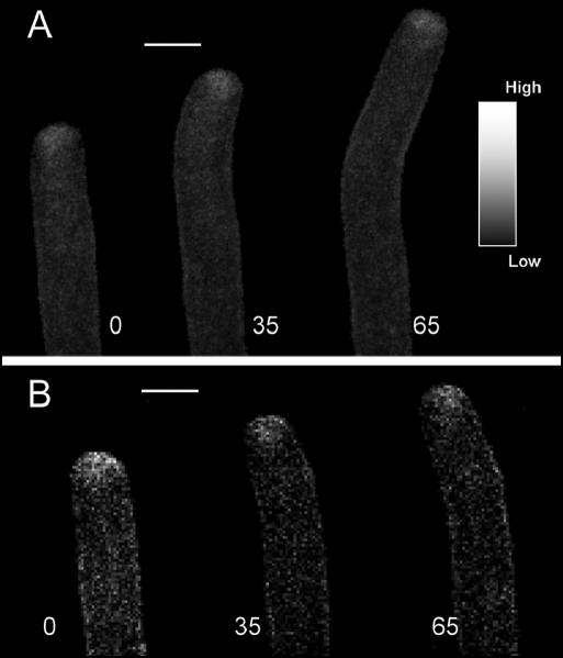
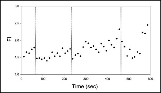
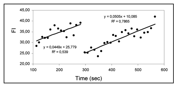
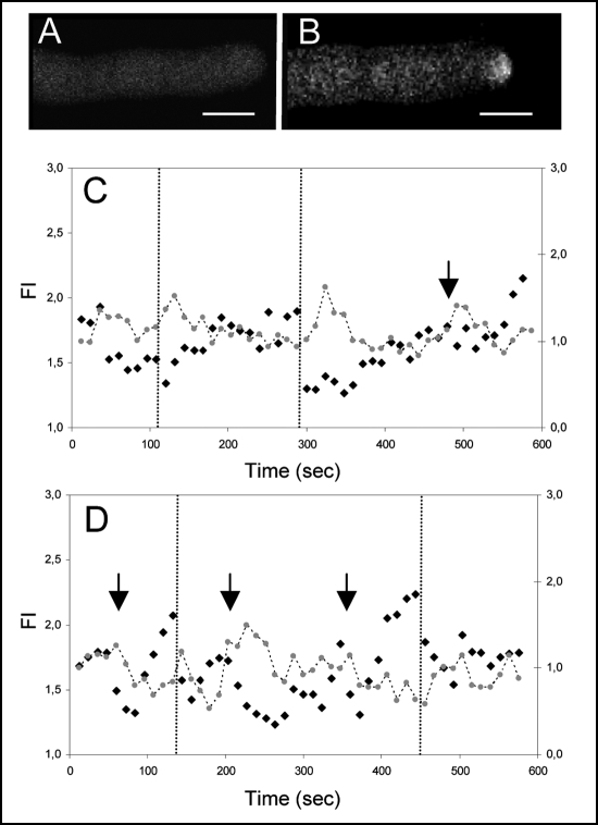
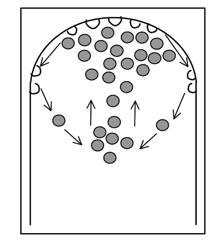
The maintenance of a cytosolic free calcium gradient (Ca(2+)](c)) and vesicle secretion in the apex of pollen tubes is essential for growth. It has been postulated that high [Ca(2+)](c) levels promote and confine vesicle fusion with the apical plasma membrane and in this study we performed a correlative analysis of both events using specific fluorescent dyes and confocal scanning microscopy. [Ca(2+)](c) was imaged with Calcium Green-1 10 kDa dextran (CG-1) while secretory events were followed with FM1-43 or FM4-64 in pollen tubes undergoing normal growth and reorientation events.During straight growth (no modification in direction), we found that changes in apical [Ca(2+)](c) accompany changes in apical FM fluorescence indicating a tight coupling between [Ca(2+)](c) and apical secretion. This coupling seems however to be perturbed during periods of reorientation of the pollen tube growth axis. Analysis of apical and sub-apical fluorescent signals during the reorientation events and subsequent re-entry in straight growth indicate that the increase in secretory events (higher fusion rate) precede the increases in [Ca(2+)](c) that should be required for the transduction of the signal.Based on these findings, we discuss a model for membrane secretion and recycling which considers the apical and sub-apical region as a functional area containing all the elements required to promote and sustain growth.

Keywords: FM1–43; [Ca2+]c; cFM4–64; endocytosis; exocytosis; secretion; tip growth.


Figures

Figure 1
Figure 1
Time course confocal images of A. umbellatus pollen tube labelled with FM dyes undergoing reorientation of the growth axis. (A) FM1–43. (B) FM4–64. With both dyes, the typical apical hotspot is visible. During changes in the growth axis, asymmetric distribution of fluorescence within the apical dome can be measured. The times (sec) at which the images were taken are indicated next to the images. Scale bar = 10 µm.
Figure 2
Figure 2
Typical variation of the FM4–64 fluorescent signal in a growing pollen tube. Since FM signal is a combination of new internalysed dye and movement of previously stained vesicles, fluorescence intensity (FI) was represented as the ratio apical/sub-apical signal (measured respectively in the first 10 µm and 10–20 µm of an individual A. umbellatus pollen tube apex). Equivalent data were obtained in 8 independent experiments. Associated to changes in the growth axis (indicated by vertical dashed lines), one can observe rapid decreases (“collapse event”) of apical/sub-apical FM signal followed by slower recovery periods where growth is approximately straight.
Figure 3
Figure 3
Linear regression analysis (solid line) of FM4–64 apical fluorescent signal (dots) experiencing a change in growth axis at sec 300. A linear pattern can be identified during straight growth modes. The collapse event that precedes reorientation interrupts the linear pattern.
Figure 4
Figure 4
Fluorescence imaging of the FM4–64 and Calcium Green-1 apical staining in a pollen tube experiencing changes in growth direction. (A) Confocal image of the CG-1 distribution. (B) Confocal image of the FM4–64 distribution. Scale bar = 10 µm. (C) Numerical representation of FM 4–64 (black dots, Y1 axis) and CG-1 (Grey dots, Y2 axis) apical fluorescence over time. Vertical lines represent changes in the pollen tube growth axis. The collapse events of FM signal precede such changes but distinct elevations of [Ca2+]c are only visible afterwards. Fluorescence intensity (FI) was measured as in Figure 2. Equivalent data were obtained in 16 independent experiments.(D) Numerical representation of FM 4–64 (black dots) and CG-1 (Grey dots) apical fluorescence over time. Measurements and symbols as in C. At ∼500 sec, a peak in [Ca2+]c could be observed without a correspondent collapse event of the FM signal; in such cases, reorientation of the growth axis was not evident.
Figure 5
Figure 5
Diagram representing the movement of labelled vesicles (grey) towards the apical region, subsequent fusion with the apical plasma membrane and membrane recycling. This cycle of secretion and recycling gives rise to the typical inverted cone shape visible with FM dyes.

References

    1. Malhó R, Castanho Coelho P, Pierson E, Derksen J. Endocytosis and membrane recycling in pollen tubes. In: Samaj J, Baluska F, Menzel D, editors. The Plant Endocytosis. Germany: Springer-Verlag; 2005. pp. 277–291.
    1. Monteiro D, Liu Q, Lisboa S, Scherer GEF, Quader H, Malhó R. Phosphoinositides and phosphatidic acid regulate pollen tube growth and reorientation through modulation of [Ca2+]c and membrane secretion. J Exp Bot. 2005;56:1665–1674. - PubMed
    1. Pierson ES, Miller DD, Callaham DA, Shipley AM, Rivers BA, Cresti M, Hepler PK. Pollen tube growth is coupled to the extracellular calcium ion flux and the intracellular calcium gradient: Effect of BAPTA-type buffers and hypertonic media. Plant Cell. 1994;6:1815–1828. - PMC - PubMed
    1. Rathore KS, Cork RJ, Robinson KR. A cytoplasmic gradient of Ca2+ is correlated with the growth of Lily pollen tubes. Develop Biol. 1991;148:612–619. - PubMed
    1. Malhó R, Trewavas A. Localized apical increases of cytosolic free calcium control pollen tube orientation. Plant Cell. 1996;8:1935–1949. - PMC - PubMed

LinkOut - more resources