. 2009 Jun 12;4(6):e5889.
doi: 10.1371/journal.pone.0005889.

MicroRNA expression profiling in mild asthmatic human airways and effect of corticosteroid therapy

Affiliations

MicroRNA expression profiling in mild asthmatic human airways and effect of corticosteroid therapy

Andrew E Williams et al. PLoS One. .

Abstract

Background: Asthma is a common disease characterised by reversible airflow obstruction, bronchial hyperresponsiveness and chronic inflammation, which is commonly treated using corticosteroids such as budesonide. MicroRNAs (miRNAs) are a recently identified family of non-protein encoding genes that regulate protein translation by a mechanism entitled RNA interference. Previous studies have shown lung-specific miRNA expression profiles, although their importance in regulating gene expression is unresolved. We determined whether miRNA expression was differentially expressed in mild asthma and the effect of corticosteroid treatment.

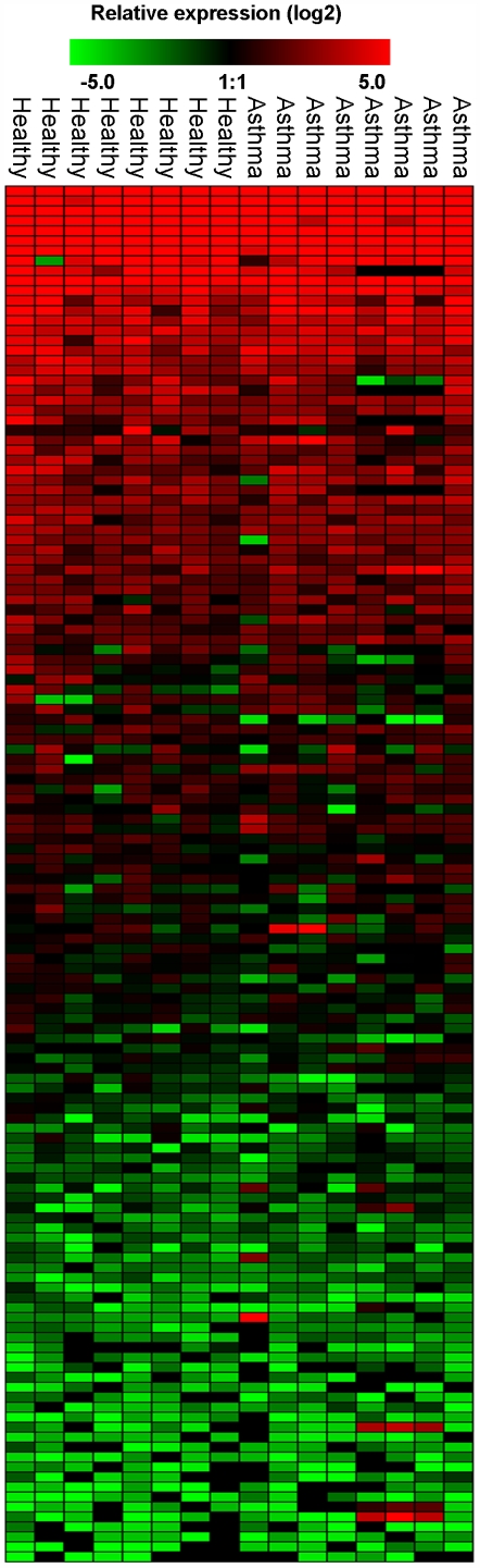
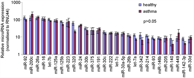
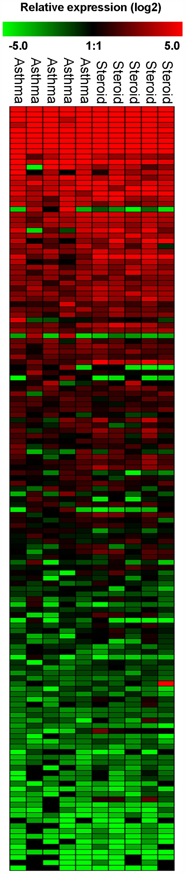
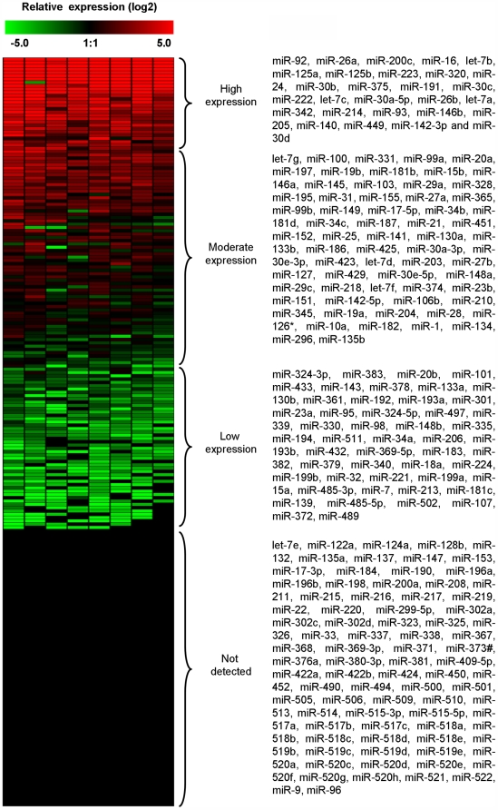
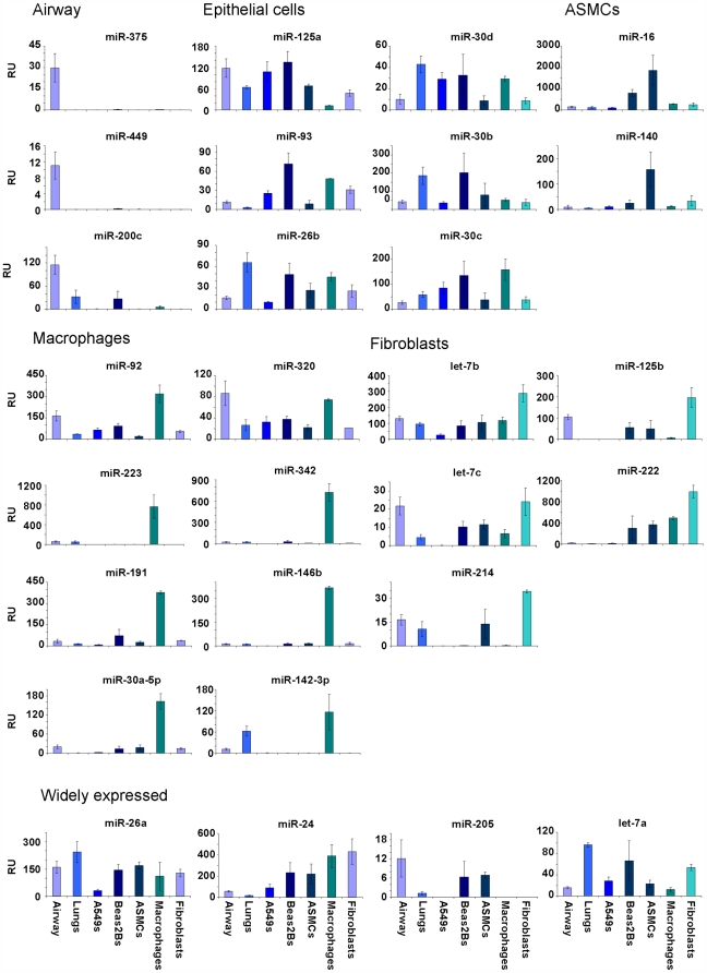
Methodology/principal findings: We have examined changes in miRNA using a highly sensitive RT-PCR based approach to measure the expression of 227 miRNAs in airway biopsies obtained from normal and mild asthmatic patients. We have also determined whether the anti-inflammatory action of corticosteroids are mediated through miRNAs by determining the profile of miRNA expression in mild asthmatics, before and following 1 month twice daily treatment with inhaled budesonide. Furthermore, we have analysed the expression of miRNAs from individual cell populations from the airway and lung. We found no significant difference in the expression of 227 miRNAs in the airway biopsies obtained from normal and mild asthmatic patients. In addition, despite improved lung function, we found no significant difference in the miRNA expression following one month treatment with the corticosteroid, budesonide. However, analysis of bronchial and alveolar epithelial cells, airway smooth muscle cells, alveolar macrophages and lung fibroblasts demonstrate a miRNA expression profile that is specific to individual cell types and demonstrates the complex cellular heterogeneity within whole tissue samples.

Conclusions: Changes in miRNA expression do not appear to be involved in the development of a mild asthmatic phenotype or in the anti-inflammatory action of the corticosteroid budesonide.


Conflict of interest statement

Competing Interests: The authors have declared that no competing interests exist.

Figures

Figure 1
Figure 1. Profile of miRNA expression in control and asthmatic patients.
A panel of 227 individual microRNAs were measured in airway biopsies obtained from normal and mild asthmatics patients using semi-quantitative real-time RT-PCR. The ΔCT method was used to calculate the relative expression (compared to the mean expression of all miRNAs measured) of each miRNA (2-(ΔCT individual miRNA – mean of 227 miRNAs)) and each sample was normalised to the snoRNA RNU44. Red indicates an increase in expression and green a decrease in expression relative to the mean expression of 227 miRNAs. Black means not detected. During the analysis, care was taken to avoid the detection of false positive, therefore ΔCT values less than 37 (that corresponded to the value of signal from the no-template control) were removed as were miRNAs that were on the limit of detectability having a sample number (n) less than n = 4. Each column represents an individual biopsy sample and each row an individual miRNA (mild asthmatic n = 8 and healthy n = 8). Statistical analysis on the sample groups was measured in Genesis using ANOVA with a p-value threshold of 0.05.
Figure 2
Figure 2. Highly expressed miRNAs did not alter in mild asthmatic airways.
Individual microRNAs were measured in airway biopsies obtained from normal and mild asthmatics patients using semi-quantitative real-time RT-PCR. The ΔCT method was used to calculate the relative expression (compared to the mean expression of all miRNAs measured) of each miRNA (2-(ΔCT individual miRNA – mean of 227 miRNAs)) and each sample was normalised to the snoRNA RNU44. A total of 28 miRNAs had a relative expression value (2-(ΔΔCT)) greater than 10 and were therefore classified as being highly expressed.
Figure 3
Figure 3. Profile of miRNA expression in asthmatic patients treated before and following treatment with budesonide.
A panel of 227 individual microRNAs were measured in airway biopsies obtained from asthmatics patients before and after 28 days treatment with twice daily inhaled budesonide (200 µg) using semi-quantitative real-time RT-PCR. The ΔCT method was used to calculate the relative expression (compared to the mean expression of all miRNAs measured) of each miRNA (2-(ΔCT individual miRNA – mean of 227 miRNAs)) and each sample was normalised to the snoRNA RNU44. Red indicates an increase in expression and green a decrease in expression relative to the mean expression of 227 miRNAs. Black means not detected. Values with a ΔCT value of 37 or greater were removed from the analysis as were miRNAs at the limit of detection having a sample number less than n = 3. Each column represents an individual biopsy sample and each row an individual miRNA (n = 5 before budesonide and n = 5 after budesonide). Statistical analysis on the sample groups was measured in Genesis using ANOVA with a p-value threshold of 0.05.
Figure 4
Figure 4. MicroRNA expression analysis of human airway biopsies.
A panel of 227 individual microRNAs were measured using semi-quantitative real-time RT-PCR on non-asthmatic, healthy human airway biopsy samples (n = 8). The ΔCT method was used to calculate the relative expression (compared to the mean expression of all miRNAs measured) of each miRNA (2-(ΔCT individual miRNA – mean of 227 miRNAs)) and each sample was normalised to the snoRNA RNU44. Red indicates an increase in expression and green a decrease in expression relative to the mean expression of 227 miRNAs.
Figure 5
Figure 5. Bioinformatic analysis of highly expressed miRNAs in human airways.
The specific miRNA targets were predicted using Targetscan 4.2. The gene targets for those miRNAs highly expressed in the airway (2-(ΔΔCT)>10). Targets that shared a high frequency across the miRNAs analysed (28 miRNAs in the highly expressed group) are plotted. Likewise, the transcription factor binding sites within 5 kb of the miRNA start site, for the same set of miRNAs, were analysed using UCSC Genome Browser. Abbreviations; MAPK, mitogen activated protein kinase; COL, collagen; EIF, eukaryotic translation initiation factor; SOCS, suppressor of cytokine signalling; IGF, insulin-like growth factor; PCDH, protocadherin.
Figure 6
Figure 6. Differential expression of highly expressed miRNAs in individual airway and lung cell populations.
The expression of miRNAs that had an expression value 10-fold or more above the average miRNA expressed in the airway, were analysed in airway biopsies, whole lung samples, alveolar epithelial cells (A549), bronchial epithelial cells (Beas2B), airway smooth muscle (ASM), alveolar macrophages (Macs) and bronchial fibroblasts (Fibs). The relative expression of each miRNA was calculated using the ΔΔCT method (calibrated to the mean value of each miRNA in the airway) generating a fold-difference in expression compared to the airways (relative expression, RU). The expression of each miRNA was attributed a grouping according to each cell population (epithelial cells (A549s or Beas2Bs), airway smooth muscle cells (ASMCs), alveolar macrophages or bronchial fibroblasts) or to whole airways or to a group having a wide distribution.

References

    1. Gregory RI, Yan KP, Amuthan G, Chendrimada T, Doratotaj B, et al. The Microprocessor complex mediates the genesis of microRNAs. Nature. 2004;432:235–240. - PubMed
    1. Denli AM, Tops BB, Plasterk RH, Ketting RF, Hannon GJ. Processing of primary microRNAs by the Microprocessor complex. Nature. 2004;432:231–235. - PubMed
    1. Yi R, Qin Y, Macara IG, Cullen BR. Exportin-5 mediates the nuclear export of pre-microRNAs and short hairpin RNAs. Genes Dev. 2003;17:3011–3016. - PMC - PubMed
    1. Hutvagner G, McLachlan J, Pasquinelli AE, Balint E, Tuschl T, et al. A cellular function for the RNA-interference enzyme Dicer in the maturation of the let-7 small temporal RNA. Science. 2001;293:834–838. - PubMed
    1. Tang G. siRNA and miRNA: an insight into RISCs. Trends Biochem Sci. 2005;30:106–114. - PubMed

Publication types