. 2009 Sep;27(9):2209-19.
doi: 10.1002/stem.150.

Circulating osteogenic precursor cells in heterotopic bone formation

Affiliations

Circulating osteogenic precursor cells in heterotopic bone formation

Robin K Suda et al. Stem Cells. 2009 Sep.

Abstract

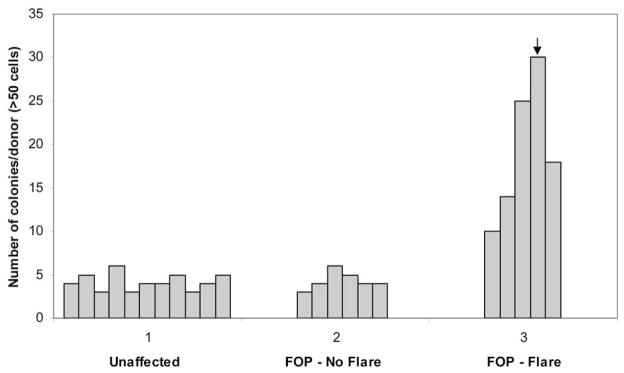
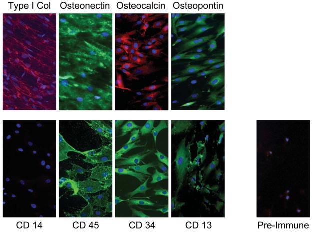
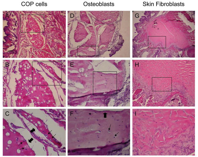
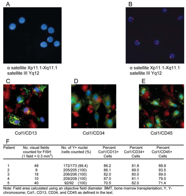
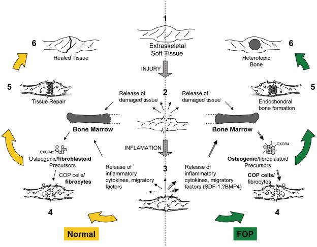
Cells with osteogenic potential can be found in a variety of tissues. Here we show that circulating osteogenic precursor (COP) cells, a bone marrow-derived type I collagen+/CD45+ subpopulation of mononuclear adherent cells, are present in early preosseous fibroproliferative lesions in patients with fibrodysplasia ossificans progressiva (FOP) and nucleate heterotopic ossification (HO) in a murine in vivo implantation assay. Blood samples from patients with FOP with active episodes of HO contain significantly higher numbers of clonally derived COP cell colonies than patients with stable disease or unaffected individuals. The highest level of COP cells was found in a patient just before the clinical onset of an HO exacerbation. Our studies show that even COP cells derived from an unaffected individual can contribute to HO in genetically susceptible host tissue. The possibility that circulating, hematopoietic-derived cells with osteogenic potential can seed inflammatory sites has tremendous implications and, to our knowledge, represents the first example of their involvement in clinical HO. Thus, bone formation is not limited to cells of the mesenchymal lineage, and circulating cells of hematopoietic origin can also serve as osteogenic precursors at remote sites of tissue inflammation.


Conflict of interest statement

Disclosures: The authors indicate no potential conflicts of interest.

Figures

Figure 1
Figure 1. Mononuclear blood-derived adherent cell (BdAC) colonies are abundant in FOP patients with recent disease activity
Blood-derived adherent cells (BdACs) were isolated from unaffected individuals (n=11), those with stable FOP (FOP – No flare, n=6), or those with active disease progression and HO (FOP - Flare, n=5). Blood samples were derived from subjects in a blinded fashion prior to isolation as a plastic-adherent, mononuclear fraction after Ficoll-gradient centrifugation. Equal numbers of mononuclear cells from each group were cultured before analysis of BdAC colonies. Shown is the number of BdAC colonies/10 cm2 derived from individuals within each group. The arrow indicates the number of colonies derived from a patient whose FOP exacerbation followed BdAC isolation by one week.
Figure 2
Figure 2. Clonally-expanded blood-derived adherent cells (BdACs) express osteoblastic and hematopoietic markers
Osteoblastic (top panels) and hematopoietic (bottom panels, left) markers detected by immunofluorescence in clonally-expanded BdACs are shown. Nuclei were counterstained with 4,6 diamidino-2-phenyindole (DAPI) and appear blue. Background immunofluorescence using pre-immune serum is also shown. Type 1 col, type 1 collagen. Original magnifcation is 200X.
Figure 3
Figure 3. Circulating osteogenic precursor (COP) cells form bone in vivo
COP cells, normal human osteoblasts, and human skin fibroblasts were transplanted subcutaneously into nude mice, and implants were excised after 8 weeks, fixed in formalin, and processed for thin-sectioning and staining with hematoxylin and eosin. Tissue sections of implants of COP cells (A–C), osteoblasts (D–F), and skin fibroblasts (G–I) are shown at an original magnification of 25X (A, D, G), 100X (B, E, H), and 200X (C, F, I). Boxes indicate regions magnified on next lower vertical panels. Thin arrows indicate osteocytes in lacune and block arrows indicate surfaces of bone-lining cells.
Figure 4
Figure 4. COP cells are derived from bone marrow
A–B, Detection of X (red/orange) and Y (green) chromosomes in COP cell interphase and metaphase nuclei from two (A, B) female patients who underwent sex-mismatched bone marrow transplants. Directly labeled fluorescent DNA probes were specific for the AT rich alpha satellite DNA sequence at the centromeric region of chromosome X (Xp11.1-Xq11.1) and for the satellite III DNA at the Yq12 region of chromosome Y. Original magnification 400X. (C–E) Identification of blood-derived adherent cells and COP cells in female sex-mismatched BMT recipients. Positive cells were identified by double-labeling immunofluorescence using specific antibodies against type I collagen and the indicated hematopoietic marker [Green, hematopoietic marker; Red, type I collagen; Blue, DAPI; Yellow/Orange, merged]. (F) Quantitation of COP cells in female sex-mismatched BMT recipients by three different combinations of markers.
Figure 5
Figure 5. COP cells are present in heterotopic bone
Lesional tissue from an FOP flare was obtained after removal from a patient’s back for suspected malignancy. COP cells, expressing type I collagen and CD 45, can be detected in pre-osseous regions of FOP lesions by in situ immunofluorescence. Original magnification is 100X. Green, type I collagen; Red, CD45; Blue, DAPI
Figure 6
Figure 6. Hypothetical mechanism of heterotopic ossification in FOP
1–3, Extraskeletal soft tissue, usually muscle, is injured causing the release of damaged tissue, inflammatory cytokines and migratory factors. SDF-1 and BMP4 are potential candidates given their roles in tissue injury and chemotaxis. 3–4, These factors may recruit circulating mesenchymal precursors of hematopoietic origin, including fibroblastoid and osteogenic precursors, which may be overexpressed or dysregulated in FOP (=▶). Circulating osteogenic precursor (COP) cells abundant in FOP patients with recent flares (=▶) home to sites of injury, perhaps through the expression of CXCR4. 5–6, Heterotopic bone formation proceeds through an endochondral process. In unaffected individuals, fibroblastoid precursors likely contribute to tissue repair and minor scar formation.

References

    1. Huss R, Lange C, Weissinger EM, Kolb HJTK. Evidence of peripheral blood-derived, plastic-adherent CD34(−/low) hematopoietic stem cell clones with mesenchymal stem cell characteristics. Stem Cells. 2000;18:252–260. - PubMed
    1. Roufosse CA, Direkze NC, Otto WR, Wright NA. Circulating mesenchymal stem cells. The International Journal of Biochemistry & Cell Biology. 2004;36:585–597. - PubMed
    1. Kuwana M, Okazaki Y, Kodama H, Izumi K, Yasuoka H, Ogawa Y, Kawakami Y, Ikeda Y. Human circulating CD14+ monocytes as a source of progenitors that exhibit mesenchymal cell differentiation. J Leukoc Biol. 2003;74:833–45. - PubMed
    1. Lee OK, Kuo TK, Chen WM, Lee KD, Hsieh SL, Chen TH. Isolation of multipotent mesenchymal stem cells from umbilical cord blood. Blood. 2004;103:1669–1675. - PubMed
    1. Zvaifler NJ, Marinova-Mutafchieva L, Adams G, Edwards CJ, Moss J, Burger JA, Maini RN. Mesenchymal precursor cells in the blood of normal individuals. Arthritis Research. 2000;2:477–488. - PMC - PubMed

Publication types

MeSH terms