. 2009 Sep;27(9):2103-13.
doi: 10.1002/stem.147.

Generation of monoclonal antibodies specific for cell surface molecules expressed on early mouse endoderm

Affiliations

Generation of monoclonal antibodies specific for cell surface molecules expressed on early mouse endoderm

Paul Gadue et al. Stem Cells. 2009 Sep.

Abstract

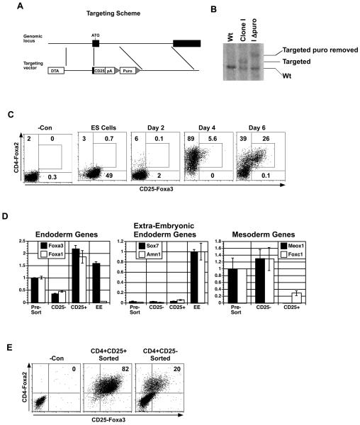
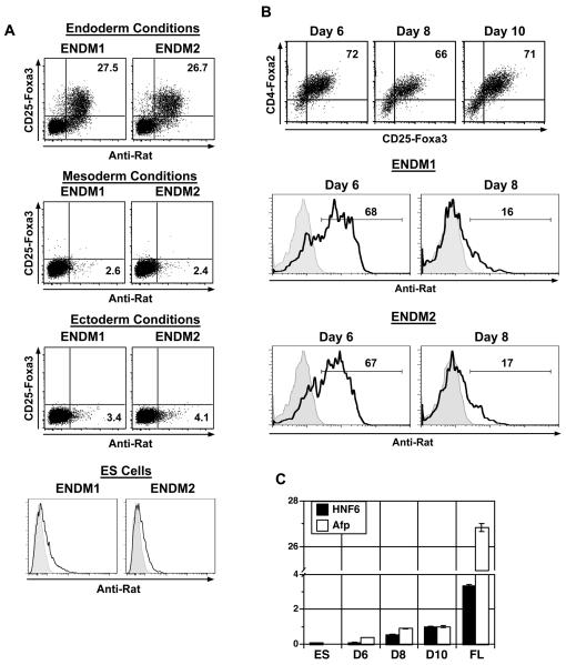
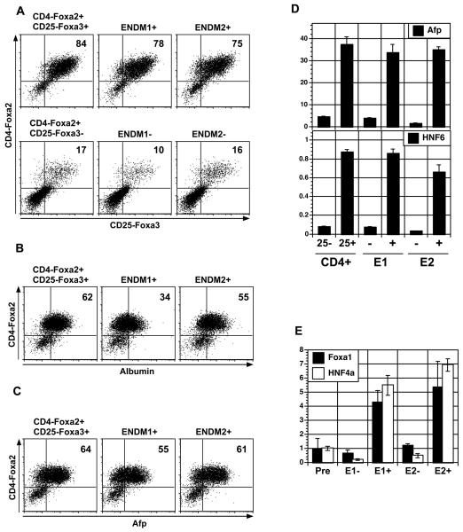
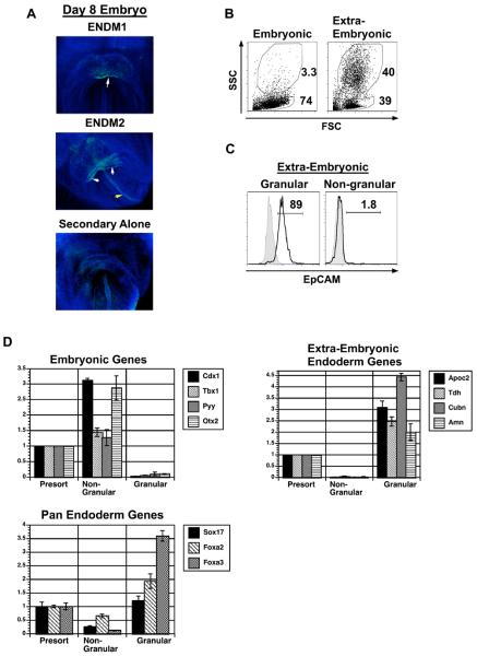
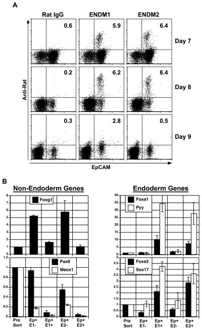
The development of functional cell populations such as hepatocytes and pancreatic beta cells from embryonic stem cell (ESC) is dependent on the efficient induction of definitive endoderm early in the differentiation process. To monitor definitive endoderm formation in mouse ESC differentiation cultures in a quantitative fashion, we generated a reporter cell line that expresses human CD25 from the Foxa3 locus and human CD4 from the Foxa2 locus. Induction of these reporter ESCs with high concentrations of activin A led to the development of a CD25-Foxa3+CD4-Foxa2+ population within 4-5 days of culture. Isolation and characterization of this population showed that it consists predominantly of definitive endoderm that is able to undergo hepatic specification under the appropriate conditions. To develop reagents that can be used for studies on endoderm development from unmanipulated ESCs, from induced pluripotent stem cells, and from the mouse embryo, we generated monoclonal antibodies against the CD25-Foxa3+CD4-Foxa2+ population. With this approach, we identified two antibodies that react specifically with endoderm from ESC cultures and from the early embryo. The specificity of these antibodies enables one to quantitatively monitor endoderm development in ESC differentiation cultures, to study endoderm formation in the embryo, and to isolate pure populations of culture- or embryo-derived endodermal cells.


Figures

Figure 1
Figure 1. Generation and characterization of the CD25-Foxa3 reporter ES cell line
(A) Schematic of Foxa3 targeting vector. (B) Southern blot analyses identifying a targeted clone and a targeted clone following removal of the selection cassette (I delta puro). (C) Kinetic analyses of CD4-Foxa2 and CD25-Foxa3 expression during endoderm formation in EBs generated from the CD25-Foxa3 reporter cell line. (D) QPCR-based gene expression analysis of CD4+CD25+ and CD4+CD25 populations isolated from day six EBs. Extra-embryonic tissue from day 8.25 mouse embryos was used as a control. Relative expression is shown with the presort population set to 1 except for the extra-embryonic endoderm genes where EE was set to 1. (E) Expression of CD4-Foxa2 and CD25-Foxa3 on CD4+CD25 and CD4+CD25+-derived populations following 12 days of culture in hepatic inducing conditions. One of three independent experiments is shown (C-E). Abbreviations: DTA, diphtheria toxin-A gene; pA, bovine poly-adenylation sequence; Puro, puromycin resistance cassette; −Con, negative control; CD25−, CD4+CD25 population; CD25+, CD4+CD25+ population; EE, extra-embryonic tissue.
Figure 2
Figure 2. Analysis of endoderm specific monoclonal antibodies on ES cell cultures
(A) Staining patterns of CD25-Foxa3, ENDM1 and ENDM2 on ES cell-derived endoderm, mesoderm, ectoderm and on undifferentiated ES cells. EBs differentiated in various conditions for 6 days were analyzed. (B) Staining patterns of CD4-Foxa2 versus CD25-Foxa3, ENDM1 and ENDM2 on sorted endoderm cells cultured in hepatocyte conditions for 1 day (day 6 total), 3 days (day 8 total) or 5 days (day 10 total). Histograms shown are gated on CD4-Foxa2+ CD25-Foxa3+ cells. (C) Afp and HNF6 expression (QPCR) in cells cultured as described in B. Relative expression is shown with the D10 population set to 1. Abbreviations: ES, ES cells; FL, day 15 fetal liver; D6, Day 6; D8, Day 8; D10, Day 10.
Figure 3
Figure 3. Isolation and characterization of ES cell-derived ENDM1+ and ENDM2+ cells
(A) CD4-Foxa2 versus CD25-Foxa3 expression on CD4+CD25+, CD4+CD25, ENDM1+, ENDM1, ENDM2+, and ENDM2 derived cells following 10 days of culture in hepatocyte inducing conditions. Populations were isolated from day 5 EBs induced under endoderm conditions. (B) Intracellular flow cytometry measuring the proportion of CD4-Foxa2+ and albumin expressing cells in the CD4+CD25+, ENDM1+ and ENDM2+-derived populations shown in A. (C) Intracellular flow cytometry measuring the proportion of CD4-Foxa2+ and Afp expressing cells in the CD4+CD25+, ENDM1+ and ENDM2+-derived populations shown in A. (D) Afp and HNF6 expression in the different populations described in A by QPCR. (E) Expression of Foxa1 and HNF4a in ENDM1+, ENDM1, ENDM2+, and ENDM2 populations sorted from 5-day-old EBs generated from E14 wild type ES cells differentiated in endoderm inducing conditions (QPCR). Relative expression is shown with the presort population set to 1. Abbreviations: CD4, CD4-Foxa2; 25, CD25-Foxa3; E1, ENDM1; E2, ENDM2; Pre, presort.
Figure 4
Figure 4. Whole mount staining of ENDM1 and ENDM2 and isolation of extra-embryonic endoderm by the side scatter flow cytometry parameter
(A) Whole mount staining analyses of day 8.25 mouse embryos showing ENDM1 and ENDM2 positive populations. White arrow indicates positive ventral endoderm and yellow arrow indicates dorsal endoderm. Embryos were co-stained with DAPI. (B) Analyses of forward and side scatter properties of the embryonic- and extra-embryonic-dissected tissues isolated form E 8.25 embryos. (C) EpCAM expression on SSChigh (granular) and SSClow (non-granular) populations from the extra-embryonic region of the embryo. Filled histogram is staining control, open histogram is EpCAM staining. (D) Gene expression analysis of the SSChigh and SSClow populations isolated from pooled whole day 8.25 mouse embryos. Populations were isolated by cell sorting based on forward and side scatter using the gates shown in B. CDNA was generated and expression of the indicated genes analyzed by QPCR. Relative expression is shown with the presort population set to 1. Data shown is the average from 3 independent experiments (60-80 embryos per experiment). Abbreviations: SSC, side scatter; FSC, forward scatter.
Figure 5
Figure 5. Expression of ENDM1 and ENDM2 during mouse embryogenesis and endoderm isolation ex vivo by cell sorting
(A) Expression of EpCAM, ENDM1 and ENDM2 on cells from embryos of indicated ages. Plots shown are gated on SSClow, non-granular cells to exclude extra-embryonic endoderm. (B) QPCR-based expression analyses of EpCAM+ ENDM1, EpCAM+ ENDM1+, EpCAM+ ENDM2, and EpCAM+ ENDM2+ populations isolated from day 8.25 mouse embryos. Cells were isolated by cell sorting, with gating on SSClow, non-granular cells to exclude extra-embryonic endoderm. Relative expression is shown with the presort population set to 1. Abbreviations: Ep, EpCAM; E1, ENDM1; E2, ENDM2.

References

    1. Conlon FL, Lyons KM, Takaesu N, et al. A primary requirement for nodal in the formation and maintenance of the primitive streak in the mouse. Development. 1994;120(7):1919–1928. - PubMed
    1. Kubo A, Shinozaki K, Shannon JM, et al. Development of definitive endoderm from embryonic stem cells in culture. Development. 2004;131(7):1651–1662. - PubMed
    1. Tada S, Era T, Furusawa C, et al. Characterization of mesendoderm: a diverging point of the definitive endoderm and mesoderm in embryonic stem cell differentiation culture. Development. 2005;132(19):4363–4374. - PubMed
    1. Gadue P, Huber TL, Paddison PJ, et al. Wnt and TGF-beta signaling are required for the induction of an in vitro model of primitive streak formation using embryonic stem cells. Proc Natl Acad Sci USA. 2006;103(45):16806–16811. - PMC - PubMed
    1. D'Amour KA, Agulnick AD, Eliazer S, et al. Efficient differentiation of human embryonic stem cells to definitive endoderm. Nat Biotechnol. 2005;23(12):1534–1541. - PubMed

Publication types

MeSH terms