

Ligation-assisted endoscopic mucosal resection of gastric heterotopic pancreas
- PMID: 19522034
- PMCID: PMC2695899
- DOI: 10.3748/wjg.15.2805
Ligation-assisted endoscopic mucosal resection of gastric heterotopic pancreas
Abstract
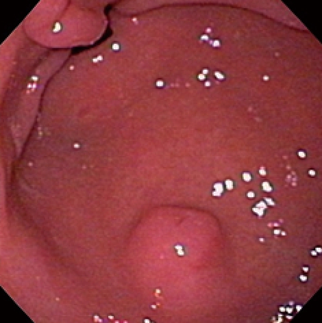
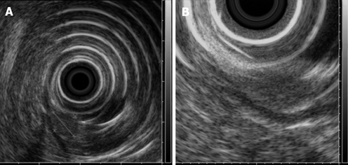
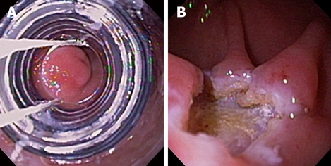
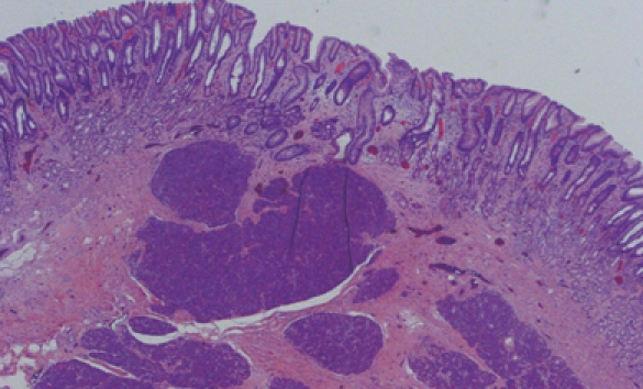
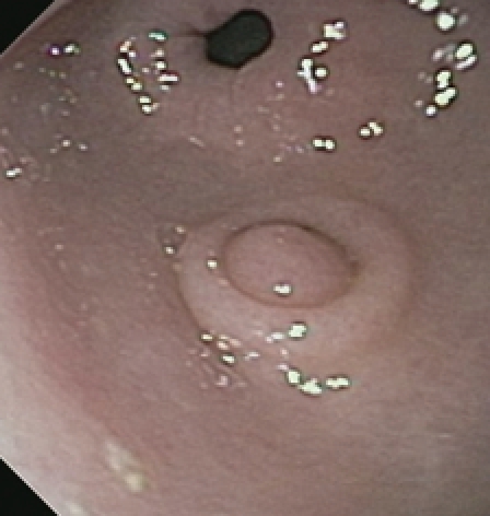
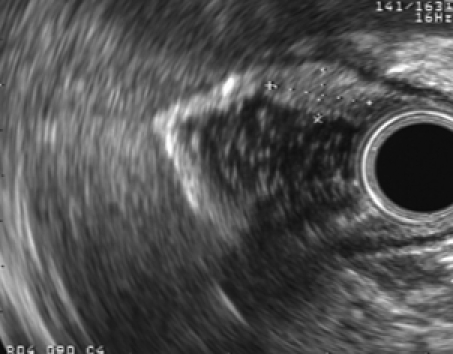
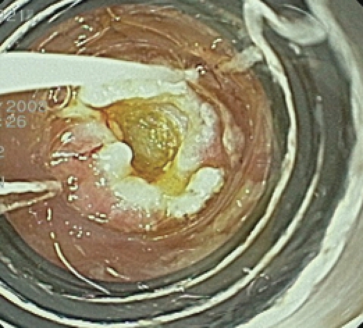
Heterotopic pancreas is a congenital anomaly characterized by ectopic pancreatic tissue. Treatment of heterotopic pancreas may include expectant observation, endoscopic resection or surgery. The aim of this report was to describe the technique of ligation-assisted endoscopic mucosal resection (EMR) for resection of heterotopic pancreas of the stomach. Two patients (both female, mean age 32 years) were referred for management of gastric subepithelial tumors. Endoscopic ultrasound in both disclosed small hypoechoic masses in the mucosa and submucosa. Band ligation-assisted EMR was performed in both cases without complications. Pathology from the resected tumors revealed heterotopic pancreas arising from the submucosa. Margins were free of pancreatic tissue. Ligation-assisted EMR is technically feasible and may be considered for the endoscopic management of heterotopic pancreas.
Figures
Similar articles
-
Endoscopic mucosal resection/endoscopic submucosal dissection for gastric heterotopic pancreas.Turk J Gastroenterol. 2013;24(4):322-9. Turk J Gastroenterol. 2013. PMID: 24254263
-
Endoscopic mucosal resection for treatment of heterotopic pancreas in the stomach.J Formos Med Assoc. 1999 Sep;98(9):643-5. J Formos Med Assoc. 1999. PMID: 10560242
-
[Ectopic pancreas in the stomach of infrequent localization in the muscularis mucosae].Rev Gastroenterol Mex. 2011 Jan-Mar;76(1):73-8. Rev Gastroenterol Mex. 2011. PMID: 21592911 Spanish.
-
Endoscopic submucosal dissection combined surgery for the treatment of ectopic gastric mucosa and ectopic pancreas in muscularis propria and serosal layer of the stomach: A rare case report and review of the literature.Medicine (Baltimore). 2025 Feb 28;104(9):e41297. doi: 10.1097/MD.0000000000041297. Medicine (Baltimore). 2025. PMID: 40020126 Free PMC article. Review.
-
Gastrointestinal: Intraductal papillary mucinous neoplasm occurring in the heterotopic pancreas of the stomach.J Gastroenterol Hepatol. 2021 Sep;36(9):2333. doi: 10.1111/jgh.15418. Epub 2021 Feb 23. J Gastroenterol Hepatol. 2021. PMID: 33624344 Review. No abstract available.
Cited by
-
Gastric heterotopic pancreas masquerading as a stromal tumor: A case report.Oncol Lett. 2015 Oct;10(4):2355-2358. doi: 10.3892/ol.2015.3593. Epub 2015 Aug 11. Oncol Lett. 2015. PMID: 26622851 Free PMC article.
-
Endoscopic submucosal dissection for gastric ectopic pancreas: a single-center experience.World J Surg Oncol. 2019 Apr 16;17(1):69. doi: 10.1186/s12957-019-1612-x. World J Surg Oncol. 2019. PMID: 30992068 Free PMC article. Clinical Trial.
-
Submucosal Tunneling Endoscopic Resection for the Management of Heterotopic Pancreas With Cystic Degeneration.ACG Case Rep J. 2020 Jul 9;7(7):e00419. doi: 10.14309/crj.0000000000000419. eCollection 2020 Jul. ACG Case Rep J. 2020. PMID: 32766361 Free PMC article.
-
Endoscopic submucosal dissection of gastric ectopic pancreas.Wideochir Inne Tech Maloinwazyjne. 2013 Sep;8(3):249-52. doi: 10.5114/wiitm.2011.33709. Epub 2013 Mar 5. Wideochir Inne Tech Maloinwazyjne. 2013. PMID: 24130642 Free PMC article.
-
Endosonographic features of histologically proven gastric ectopic pancreas.Gastroenterol Res Pract. 2014;2014:160601. doi: 10.1155/2014/160601. Epub 2014 Oct 12. Gastroenterol Res Pract. 2014. PMID: 25371670 Free PMC article.
References
-
- Elfving G, Hästbacka J. Pancreatic heterotopia and its clinical importance. Acta Chir Scand. 1965;130:593–602. - PubMed
-
- Lai EC, Tompkins RK. Heterotopic pancreas. Review of a 26 year experience. Am J Surg. 1986;151:697–700. - PubMed
-
- Ormarsson OT, Gudmundsdottir I, Mårvik R. Diagnosis and treatment of gastric heterotopic pancreas. World J Surg. 2006;30:1682–1689. - PubMed
-
- Dolan RV, ReMine WH, Dockerty MB. The fate of heterotopic pancreatic tissue. A study of 212 cases. Arch Surg. 1974;109:762–765. - PubMed
-
- Lee TH, Wang HP, Huang SF, Wang TH, Lin JT. Endoscopic mucosal resection for treatment of heterotopic pancreas in the stomach. J Formos Med Assoc. 1999;98:643–645. - PubMed
Publication types
MeSH terms
LinkOut - more resources
Full Text Sources
