. 2009 Aug 18;48(32):7636-45.
doi: 10.1021/bi900031w.

Biochemical and structural characterization of the human TL1A ectodomain

Affiliations

Biochemical and structural characterization of the human TL1A ectodomain

Chenyang Zhan et al. Biochemistry. .

Abstract

TNF-like 1A (TL1A) is a newly described member of the TNF superfamily that is directly implicated in the pathogenesis of autoimmune diseases, including inflammatory bowel disease, atherosclerosis, and rheumatoid arthritis. We report the crystal structure of the human TL1A extracellular domain at a resolution of 2.5 A, which reveals a jelly-roll fold typical of the TNF superfamily. This structural information, in combination with complementary mutagenesis and biochemical characterization, provides insights into the binding interface and the specificity of the interactions between TL1A and the DcR3 and DR3 receptors. These studies suggest that the mode of interaction between TL1A and DcR3 differs from other characterized TNF ligand/receptor complexes. In addition, we have generated functional TL1A mutants with altered disulfide bonding capability that exhibit enhanced solution properties, which will facilitate the production of materials for future cell-based and whole animal studies. In summary, these studies provide insights into the structure and function of TL1A and provide the basis for the rational manipulation of its interactions with cognate receptors.


Figures

Figure 1
Figure 1. Sequence alignment of conventional TNF ligands
Those ligands capable of binding human DcR3 (TL1A, mouse TL1A, LIGHT, FasL) are treated as a group (upper four sequences). The remaining conventional ligands are displayed below the black line. Residues that are identical in only one of the two groups are shown in blue. Residues that are identical in both groups are shown in red. Additional conserved residues are colored green. Secondary structure of TL1A is displayed atop the alignment, with arrows representing β-strands and a cylinder representing the α-helix. The numbering is based on the sequence of human TL1A ectodomain and is consistently referenced in the text. With the exception of murine TL1A, all proteins are from Homo sapiens.
Figure 2
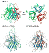
Figure 2. Ribbon structure of TL1A trimer assembly
(A) The three chains of TL1A are individually colored in cyan, green and red. The right panel is rotated by 90° around the horizontal axis relative to the left panel. The β-stands are labeled as shown on Figure 1. Each loop is referenced in the text by the two strands connected to that loop. (B and C) Superimpositions of the TL1A structure with TRAIL and CD40L. TL1A is colored in blue and gray, TRAIL is in green and CD40L is in orange.
Figure 3
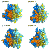
Figure 3. Different quaternary structures of TNF family members
Two protomers of each trimer assembly (TL1A, TNFα -PDB code 2TNF, CD40L -PDB code 1ALY and TRAIL -PDB code 1D4V) are shown as surface representation and the third protomer above is shown as a transparent ribbon. Hydrophobic/hydrophilic residues are colored in orange/blue in one protomer and yellow/cyan in the neighboring protomer.
Figure 4
Figure 4
(A) Gel filtration of TL1A and the DcR3/TL1A(C95S/C135S) complex. The chromatogram for TL1A alone is drawn in purple and that for the DcR3/TL1A(C95S/C135S) mixture in blue. The elution profile of the wild type TL1A/DcR3 mixture is similar to that of TL1A(C95S/C135S)/DcR3 (data not shown). The calculated molecular weights of the TL1A trimer and the DcR3 cysteine-rich domain are 62.67 kDa and 20.1 kDa, respectively. The cyan, green, purple and red arrow heads indicate the positions of standards with molecular weights of 150kDa, 66 kDa, 29kDa and 12.4 kDa, respectively. (B) Velocity sedimentation analysis of TL1A. Sedimentation coefficients of TL1A were measured at three concentrations of the protein: 10.7 μ M, 26.7 μ M and 47.3 μ M.
Figure 5
Figure 5. Disulfide bond content
(A) Non-reducing SDS-PAGE for wild type TL1A native protein (lane A), C95S (lane B) and C135S (lane C) mutants. Protein standard ladder is shown on the right side. (B and C) Models of disulfide connectivity. Each TL1A subunit is represented by a green circle, C95 and C135 by red and yellow squares, respectively. Putative disulfide bonds are shown as black sticks.
Figure 6
Figure 6. DcR3 binding determinants
A series of residues on the surface of the TL1A C95S/C135S double mutant were mutated and their binding to the DcR3 receptor was assessed by SPR. (A) The sensorgram of R99A-R103A-D108A triple mutant. Solid line: Wild type; dashed line: R99A-R103A-D108A triple mutant. (B) The sensorgram of Y121F single mutant. Solid line: Wild type; dashed line: Y121F single mutant. (C) The effects of all mutations on DcR3 binding affinity relative to the wild type. The maximum association response of each mutant is divided by that of the wild type TL1A and represented in the histogram as percentage of wild type binding response. Vertical bars on each column represent the standard errors between two replicated measurements.
Figure 7
Figure 7. Receptor binding interfaces in TNF ligands
In the upper two panels, orange patches indicate the surfaces of LTα (panel A, PDB code 1TNR) and TRAIL (panel B, PDB code 1D4V) that are involved in receptor binding based on co-crystal structures with their respective receptors (17). In the lower two panels, the residues important for the TL1A-DcR3 interaction, based on mutagenesis studies, are shown in orange as a surface representation in panel C and stick representation in panel D. The residues that may indirectly impact interaction with DcR3 through effects on protein stability or oligomeric state are shown in red.

References

    1. Migone TS, Zhang J, Luo X, Zhuang L, Chen C, Hu B, Hong JS, Perry JW, Chen SF, Zhou JX, Cho YH, Ullrich S, Kanakaraj P, Carrell J, Boyd E, Olsen HS, Hu G, Pukac L, Liu D, Ni J, Kim S, Gentz R, Feng P, Moore PA, Ruben SM, Wei P. TL1A is a TNF-like ligand for DR3 and TR6/DcR3 and functions as a T cell costimulator. Immunity. 2002;16:479–492. - PubMed
    1. Chew LJ, Pan H, Yu J, Tian S, Huang WQ, Zhang JY, Pang S, Li LY. A novel secreted splice variant of vascular endothelial cell growth inhibitor. Faseb J. 2002;16:742–744. - PubMed
    1. Yang CR, Hsieh SL, Teng CM, Ho FM, Su WL, Lin WW. Soluble decoy receptor 3 induces angiogenesis by neutralization of TL1A, a cytokine belonging to tumor necrosis factor superfamily and exhibiting angiostatic action. Cancer Res. 2004;64:1122–1129. - PubMed
    1. Bamias G, Mishina M, Nyce M, Ross WG, Kollias G, Rivera-Nieves J, Pizarro TT, Cominelli F. Role of TL1A and its receptor DR3 in two models of chronic murine ileitis. Proc Natl Acad Sci U S A. 2006;103:8441–8446. - PMC - PubMed
    1. Pappu BP, Borodovsky A, Zheng TS, Yang X, Wu P, Dong X, Weng S, Browning B, Scott ML, Ma L, Su L, Tian Q, Schneider P, Flavell RA, Dong C, Burkly LC. TL1A-DR3 interaction regulates Th17 cell function and Th17-mediated autoimmune disease. J Exp Med. 2008;205:1049–1062. - PMC - PubMed

Publication types

MeSH terms

Substances