. 2009 Jul;276(14):3729-43.
doi: 10.1111/j.1742-4658.2009.07089.x. Epub 2009 Jun 11.

Lysosomal localization of GLUT8 in the testis--the EXXXLL motif of GLUT8 is sufficient for its intracellular sorting via AP1- and AP2-mediated interaction

Affiliations
Free PMC article

Lysosomal localization of GLUT8 in the testis--the EXXXLL motif of GLUT8 is sufficient for its intracellular sorting via AP1- and AP2-mediated interaction

Muhammed Kasim Diril et al. FEBS J. 2009 Jul.
Free PMC article

Abstract

The class III sugar transport facilitator GLUT8 co-localizes with the lysosomal protein LAMP1 in heterologous expression systems. GLUT8 carries a [D/E]XXXL[L/I]-type dileucine sorting signal that has been postulated to retain the protein in an endosomal/lysosomal compartment via interactions with clathrin adaptor protein (AP) complexes. However, contradictory findings have been described regarding the subcellular localization of the endogenous GLUT8 and the adaptor proteins that interact with its dileucine motif. Here we demonstrate that endogenous GLUT8 is localized in a late endosomal/lysosomal compartment of spermatocytes and spermatids, and that the adaptor complexes AP1 and AP2, but not AP3 or AP4, interact with its N-terminal intracellular domain (NICD). In addition, fusion of the GLUT8 NICD to the tailless lumenal domain of the IL-2 receptor alpha chain (TAC) protein (interleukin-2 receptor a chain) targeted the protein to intracellular membranes, indicating that its N-terminal dileucine signal is sufficient for endosomal/lysosomal targeting of the transporter. The localization and targeting of GLUT8 show striking similarities to sorting mechanisms reported for lysosomal proteins. Therefore, we suggest a potential role for GLUT8 in the so far unexplored substrate transport across intracellular membranes.


Figures

Fig. 1
Fig. 1
Co-localization of GLUT8 with LAMP1 in mouse testis. Immunohistochemistry of paraffin-embedded testis sections from wild-type (A–I) and GLUT8-deficient mice (Slc2a8−/−) (J–L). GLUT8 was not detectable in testis from GLUT8 knockout animals (J). In testis from wild-type animals (Slc2a8+/+), GLUT8 staining (A,D) overlaps (C,F) with the lysosomal protein LAMP1 (B,E). In contrast, the Golgi marker GM130 (H) did not co-localize with GLUT8 (G), as seen by the lack of overlap between the two proteins (I). Scale bars = 10 μm.
Fig. 2
Fig. 2
The [DE]XXXL[LI] motif of GLUT8 interacts with endogenous AP1 and AP2 in GST pulldown assays. GST pulldown assays were performed using lysates of rat brain (A) and HEK293 cells (B), clathrin-coated vesicle membranes enriched from rat brains (C), and lysates from mouse testis (D). The recombinant wild-type or mutated N-terminus of GLUT8 fused to GST was used as bait. The first lane in each panel represents a control for the lysates or membranes used in the pulldown assays (percentage of the total in parentheses).
Fig. 3
Fig. 3
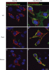
GLUT8 sorting is not altered in mocha and pearl cells lacking AP3 subunits. GLUT8 and the LL/AA mutant were overexpressed in either wild-type (WT) or AP3-deficient (pearl, mocha) mouse embryonic fibroblasts. Differential staining was performed in order to differentiate between plasma membrane and total GLUT8. Plasma membrane GLUT8 (A,C,E) or LL/AA mutant (B,D,F) was detected by incubating cells with the anti-haemagglutinin IgG in cell culture prior to fixation (in green). The haemagglutinin antibody recognizes plasma membrane GLUT8 via a haemagglutinin epitope that was introduced into the first extracellular loop of the transporter. Total GLUT8 was visualized using the C-terminal anti-GLUT8 IgG (in red) after fixation and permeabilization of cells. Scale bars = 10 μm.
Fig. 4
Fig. 4
Co-localization of GLUT8 and LAMP1 is not affected in AP3-deficient cells. GLUT8 and the LL/AA mutant were overexpressed in either wild-type (A–F) or mocha (G–L) fibroblasts. Co-localization of GLUT8 and LAMP1 is seen to be independent of the presence (A–C) or absence (G–I) of AP3. However, the GLUT8-LL/AA mutant does not co-localize with LAMP1 (F,L), but instead appears at the plasma membrane in wild-type (E) as well as mutant (K) cells. Scale bars = 10 μm.
Fig. 5
Fig. 5
GLUT8 accumulates at the plasma membrane when cells are depleted of adaptor proteins or the clathrin heavy chain. (A) HeLa cells were transfected twice within 5 days with siRNA for AP1, AP2, AP1/AP2 or the clathrin heavy chain (CHC). After the second transfection, cells were analysed for efficient protein knockdown after 48 h by western blot analysis. (B) Alexa Fluor 488-conjugated transferrin uptake or LAMP1 antibody internalization were performed as described previously [40]. AP2 and CHC knockdown dramatically affects LAMP1 and transferrin receptor trafficking, leading to accumulation of the two proteins at the plasma membrane. Knockdown of AP1 leads to a modest level of GLUT8 in plasma membrane. In contrast, GLUT8 accumulates at the plasma membrane in cells transfected with AP2 or CHC siRNA. Scale bars = 10 μm.
Fig. 6
Fig. 6
The [DE]XXXL[LI] motif of GLUT8 is sufficient for its intracellular retention. (A) Four chimeras (tailless interleukin-2 receptor α chain (TAC), a CD3-δ–TAC chimera, TAC–wild-type GLUT8 N-terminus and TAC–LL/AA-GLUT8 N-terminus) were transfected into HeLa cells. (B) Appearance of the proteins at the plasma membrane was assessed by TAC antibody internalization (labelled in green), and the overall distribution of the chimeric proteins was analysed after fixation and permeabilization of the cells (labelled in red). The tailless interleukin-2 receptor α chain construct appears at the plasma membrane only (B,a–c), whereas the CD3δt3t2–TAC chimera containing the EXXXLL consensus sequence is internalized from the membrane, as indicated by the internalized TAC antibody labelled in green (B,d). The GLUT8–TAC chimera is not targeted to the plasma membrane (green labelling in B,g). Mutating the dileucine motif of GLUT8 to LL/AA results in the opposite picture compared with the GLUT8–TAC protein, i.e. localization of the GLUT8-LL/AA–TAC chimera is restricted to the plasma membrane (B,k). Scale bars = 10 μm.

References

    1. Scheepers A, Joost HG, Schurmann A. The glucose transporter families SGLT and GLUT: molecular basis of normal and aberrant function. JPEN J Parenter Enteral Nutr. 2004;28:364–371. - PubMed
    1. Joost HG, Thorens B. The extended GLUT-family of sugar/polyol transport facilitators: nomenclature, sequence characteristics, and potential function of its novel members. Mol Membr Biol. 2001;18:247–256. - PubMed
    1. Carayannopoulos MO, Chi MM, Cui Y, Pingsterhaus JM, McKnight RA, Mueckler M, Devaskar SU, Moley KH. GLUT8 is a glucose transporter responsible for insulin-stimulated glucose uptake in the blastocyst. Proc Natl Acad Sci USA. 2000;97:7313–7318. - PMC - PubMed
    1. Augustin R, Riley J, Moley KH. GLUT8 contains a [DE]XXXL[LI] sorting motif and localizes to a late endosomal/lysosomal compartment. Traffic. 2005;6:1196–1212. - PubMed
    1. Lisinski I, Schurmann A, Joost HG, Cushman SW, Al-Hasani H. Targeting of GLUT6 (formerly GLUT9) and GLUT8 in rat adipose cells. Biochem J. 2001;358:517–522. - PMC - PubMed

Publication types

MeSH terms