. 2009 Oct;11(10):1533-49.
doi: 10.1111/j.1462-5822.2009.01345.x. Epub 2009 Jun 11.

Alternative infectious entry pathways for dengue virus serotypes into mammalian cells

Affiliations

Alternative infectious entry pathways for dengue virus serotypes into mammalian cells

Eliana G Acosta et al. Cell Microbiol. 2009 Oct.

Abstract

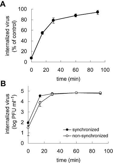
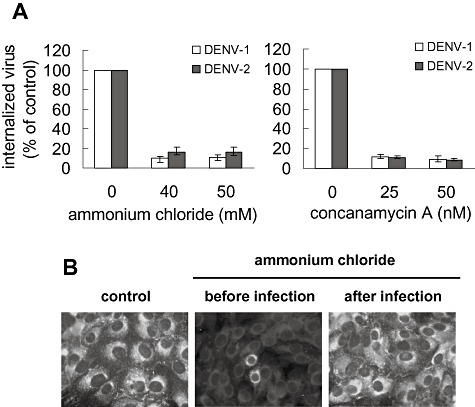
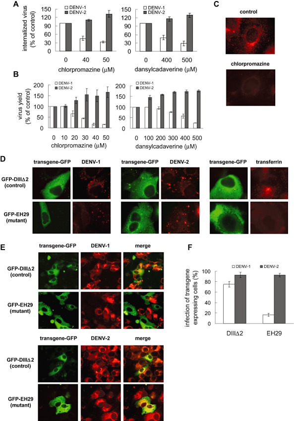
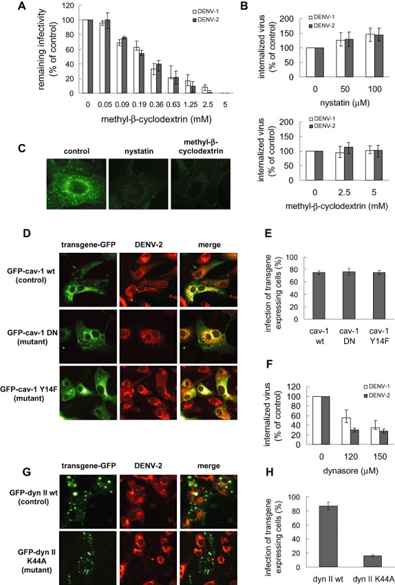
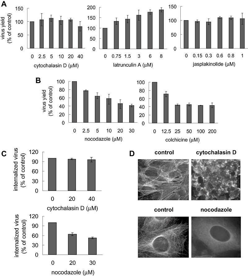
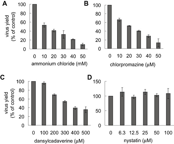
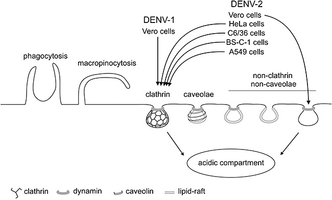
The entry of two dengue virus (DENV) serotypes into Vero cells was analysed using biochemical inhibitors, dominant negative mutants of cellular proteins involved in endocytic pathways, fluorescence microscopy and infectivity determinations. By treatment with dansylcadaverine and chlorpromazine and overexpression of a dominant negative form of the Eps15 protein, a clathrin-mediated endocytosis for productive DENV-1 internalization into Vero cells was demonstrated whereas the infectious entry of DENV-2 in the same cell system was independent of clathrin. Treatment with the inhibitors nystatin and methyl-beta-cyclodextrin, as well as transfection of Vero cells with dominant negative caveolin-1, had no effect on DENV-2 virus infection. It was also shown, by using the K44A mutant and the inhibitor dynasore, that dynamin was required for DENV-2 entry. Consequently, the infectious entry of DENV-2 into Vero cells occurs by a non-classical endocytic pathway independent of clathrin, caveolae and lipid rafts, but dependent on dynamin. By contrast, DENV-2 entry into A549 cells was clathrin-dependent, as previously reported in HeLa, C6/36 and BS-C-1 cells. Our results conclusively show, for the first time, a differential mode of infective entry for DENV-1 and DENV-2 into a common host cell, Vero cells, as well as alternative entry pathways for a given serotype, DENV-2, into different types of cells.


Figures

Figure 1
Figure 1
Kinetics and rate of DENV internalization into Vero cells.
A. Vero cells were infected with DENV‐2 during 1 h at 4°C and then transferred to 37°C. At the indicated times after incubation at 37°C, extracellular virus was inactivated with proteinase K. Results are shown as a percentage of internalized virus when compared with controls in which PBS was substituted for proteinase K.
B. Vero cells were infected with DENV‐2 at 37°C (non‐synchronized infection) or at 4°C and then transferred to 37°C (synchronized infection). At the indicated times extracellular virus was inactivated with proteinase K. Results are expressed as number of internalized PFU.
Figure 2
Figure 2
DENV‐1 and DENV‐2 entry into Vero cells is dependent of acidic pH.
A. Cells were treated with ammonium chloride or concanamycin A and infected with DENV‐1 or DENV‐2. After 1 h internalization in the presence of the drugs, extracellular virus was inactivated with proteinase K and the cell pellets were plated onto Vero cells to determine internalized virus by an infectious centre assay. Results are expressed as percentage of internalized virus with respect to a control without drug treatment. Each point shows the mean ± SD of two independent experiments.
B. Cells were treated with 50 mM ammonium chloride before or after infection with DENV‐2 or were left untreated (control). At 48 h p.i. immunostaining was carried out using mouse anti‐E glycoprotein antibody and FITC‐labelled anti‐mouse IgG.
Figure 3
Figure 3
Entry of DENV‐1 into Vero cells requires clathrin‐mediated endocytosis while entry of DENV‐2 is clathrin‐independent.
A. Cells were treated with chlorpromazine or dansylcadaverine and infected with DENV‐1 or DENV‐2. After 1 h internalization in the presence of the drugs, monolayers were treated with proteinase K and the cell pellets were plated onto Vero cells to determine internalized virus by an infectious centre assay.
B. Cells were treated as in (A) and infected with DENV‐1 or DENV‐2. Virus yields were quantified at 48 h p.i. Results are expressed as percentage of virus multiplication with respect to a control without drug treatment.
C. Cells were treated with 50 μM chlorpromazine or left untreated (control) and incubated with TRITC‐labelled transferrin.
D. Cells transiently transfected with the constructs GFP‐DIIIΔ2 or GFP‐EH29 were infected with DENV‐1 or DENV‐2 or incubated with TRITC‐labelled transferrin. After 1 h cells were fixed and internalized viral particles were visualized by immunofluorescence staining using mouse anti‐E glycoprotein antibody and TRITC‐labelled anti‐mouse IgG.
E. Cells transiently transfected with GFP‐DIIIΔ2 or GFP‐EH29 were infected with DENV‐1 or DENV‐2. After 24 h cells were fixed and viral antigen expression was visualized by immunofluorescence staining using mouse anti‐E glycoprotein antibody and TRITC‐labelled anti‐mouse IgG.
F. For quantification of samples shown in (E), 250 transfected cells with similar levels of GFP expression were screened and cells positive for viral antigen were scored.
In (A), (B) and (F) results are expressed as the mean ± SD of two independent experiments.
Figure 4
Figure 4
DENV‐2 entry into Vero cells is independent of caveolae/lipid‐rafts but is dependent on dynamin.
A. DENV‐1 or DENV‐2 suspensions were incubated at 37°C with various concentrations of methyl‐β‐cyclodextrin. After 1 h remaining infectivity was determined.
B. Vero cells were pretreated with nystatin or methyl‐β‐cyclodextrin. Then monolayers were washed with PBS and infected with DENV‐1 or DENV‐2 in culture medium without serum. After 1 h internalization, cultures were treated with proteinase K and the cell pellets were plated onto Vero cells to determine internalized virus by an infectious centre assay. Results are expressed as percentage of internalized virus with respect to a control without drug treatment.
C. Cells were untreated (control) or treated with 100 μM nystatin or 5 mM methyl‐β‐cyclodextrin. Then cultures were incubated with FITC‐labelled cholera toxin.
D. Cells transiently transfected with the constructs GFP‐cav‐1 wt, GFP cav‐1 DN or GFP‐cav‐1 Y14F were infected with DENV‐2. After 24 h infection cultures were fixed and immunofluorescence staining was performed.
E. For quantification of samples shown in (D), 250 transfected cells with similar levels of GFP expression were screened and cells positive for viral antigen were scored.
F. Vero cells were treated with dynasore, infected with DENV‐1 or DENV‐2 in the presence of the drug and then processed as in (B).
G. Cells transiently transfected with the constructs GFP‐dyn II wt or GFP‐dyn II K44A were infected with DENV‐2. After 24 h infection cultures were fixed and immunofluorescence staining was performed.
H. For quantification of samples shown in (G), 250 transfected cells with similar levels of GFP expression were screened and cells positive for viral antigen were scored.
In (A), (B), (E), (F) and (H) values represent the mean ± SD of two independent experiments.
Figure 5
Figure 5
Effect of cytoskeleton‐disrupting agents on DENV‐2 entry into Vero cells.
A and B. Cells were treated with increasing concentrations of cytochalasin D, latrunculin A, jasplakinolide, nocodazole or colchicine and infected with DENV‐2. Virus yields were determined at 48 h p.i. Results are expressed as percentage of virus multiplication with respect to a control without drug treatment. Each point shows the mean ± SD of two independent experiments.
C. Cells were treated with cytochalasin D or nocodazole and infected with DENV‐2. After 1 h internalization in the presence of the drugs, monolayers were treated with proteinase K and the cell pellets were plated onto Vero cells to determine internalized virus by an infectious centre assay.
D. Cells were untreated (control) or treated with 40 μM cytochalasin D or 30 μM nocodazole and then stained with FITC‐labelled phalloidin or mouse anti‐tubulin antibody and TRITC‐labelled anti‐mouse IgG.
Figure 6
Figure 6
DENV‐2 infection of A549 cells is dependent on clathrin‐mediated endocytosis. A549 cells were treated with various concentrations of ammonium chloride (A), chlorpromazine (B), dansylcadaverine (C) or nystatin (D) and infected with DENV‐2. Virus yields were quantified at 48 h p.i. Results are expressed as percentage of virus multiplication with respect to a control without drug treatment. Each point shows the mean ± SD of two independent experiments.
Figure 7
Figure 7
Endocytic pathways utilized by DENV‐1 and DENV‐2 to infect different cell lines. DENV‐1 infection of Vero cells occurs through a classical clathrin‐mediated dynamin‐dependent endocytosis, while infection of this same cell line by DENV‐2 takes place through a non‐classical endocytic pathway independent of clathrin, caveolin‐1 and lipid‐rafts, but dependent on dynamin. This entry route differs from the clathrin‐mediated pathway followed by DENV‐2 to infect A549 cells and, as previously shown, to infect C6/36 cells (Acosta et al., 2008b; Mosso et al., 2008), HeLa cells (Krishnan et al., 2007) and BS‐C‐1 cells (van der Schaar et al., 2008).

References

    1. Acosta, E.G. , Talarico, L.B. , and Damonte, E.B. (2008a) Cell entry of dengue virus. Future Virol 3: 471–479.
    1. Acosta, E.G. , Castilla, V. , and Damonte, E.B. (2008b) Functional entry of dengue virus into Aedes albopictus mosquito cells is dependent on clathrin‐mediated endocytosis. J Gen Virol 89: 474–484. - PubMed
    1. Aizaki, H. , Morikawa, K. , Fukasawa, M. , Hara, H. , Inoue, Y. , Tani, H. , et al. (2008) Critical role of virion‐associated cholesterol and sphingolipid in hepatitis C virus infection. J Virol 82: 5715–5724. - PMC - PubMed
    1. Altmeyer, R. (2004) Virus attachment and entry offer numerous targets for antiviral therapy. Curr Pharm Des 10: 3701–3712. - PubMed
    1. Amstutz, B. , Gastaldelli, M. , Kälin, S. , Imelli, N. , Boucke, K. , Wandeler, E. , et al. (2008) Subversion of CtBP1‐controlled macropinocytosis by human adenovirus serotype 3. EMBO J 27: 956–969. - PMC - PubMed

Publication types

LinkOut - more resources