

Internal jugular vein vascular malformation presenting as mass at root of neck: a case report
- PMID: 19523242
- PMCID: PMC2703615
- DOI: 10.1186/1472-6815-9-5
Internal jugular vein vascular malformation presenting as mass at root of neck: a case report
Abstract
Background: We report a case of vascular malformation arising from internal jugular vein presenting as mass at root of neck with no clinical stigmata which to the best of our knowledge is the first reported case of an intrinsic vascular malformation arising from the internal jugular vein. Magnetic resonance imaging features of this new entity have been described.
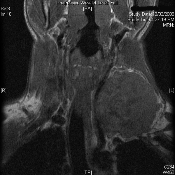
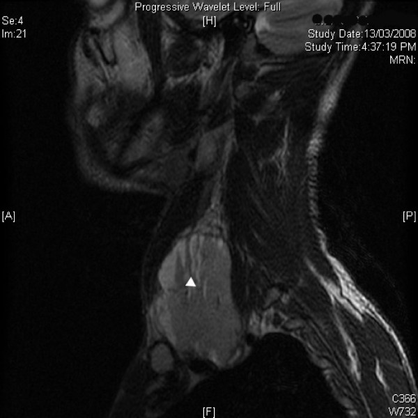
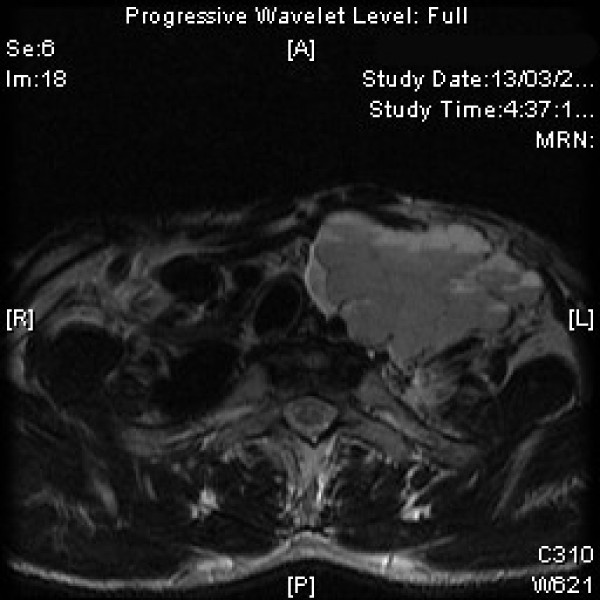
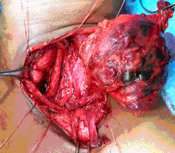
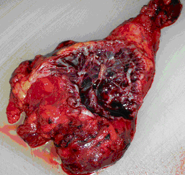
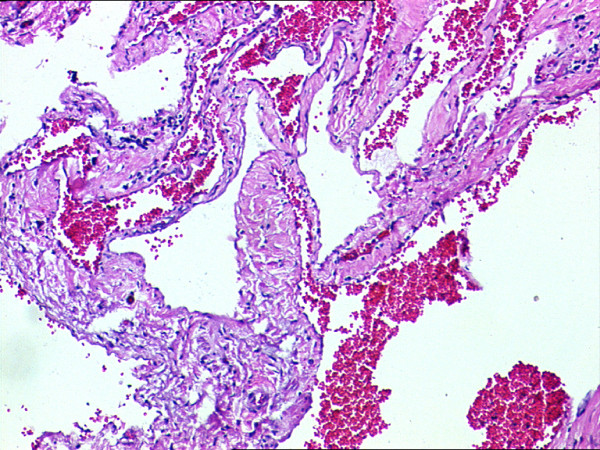
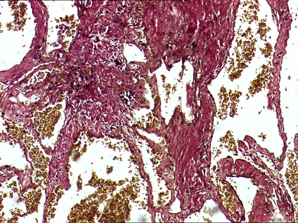
Case presentation: A 27 year male presented with a gradually enlarging, asymptomatic swelling on left supraclavicular region with normal overlying skin. A soft mass, about 7 x 7 cm with restricted mobility was found with normal cranial nerve function. Fine needle aspiration cytology showed a hemorrhagic aspirate. Doppler showed a mass displacing left carotid artery posteriorly while left internal jugular vein was not visualized. Magnetic resonance imaging showed a well defined mass isointense to hypointense on T1 weighted and hyperintense on T2 weighted and STIR images with fluid-fluid levels. On exploration, a vascular mass arising from left internal jugular vein was found with good tissue planes, which was excised after ligating the patent internal jugular vein above and below the lesion. Histopathologic examination confirmed the diagnosis of vascular malformation.
Conclusion: The diagnosis of intrinsic vascular malformation arising from internal jugular vein should be kept in differential while dealing with masses at root of neck and magnetic resonance imaging features may help in the pre-operative diagnosis of this entity.
Figures
References
-
- Mulliken JB, Glowacki J. Hemangiomas and vascular malformation in infants and children: a classification based on endothelial characteristics. Plast Reconstr Surg. 1982;69:412–422. - PubMed
-
- Sarteschi LM, Bonanomi G, Mosca F, Ferrari M. External jugular vein hemangioma occurring as a lateral neck mass. J Ultrasound Med. 1999;18:719–721. - PubMed
-
- Finn MC, Glowacki J, Mulliken JB. Congenital vascular lesions: clinical applications of a new classification. J Pediatr Surg. 2004;18:894–900. - PubMed
LinkOut - more resources
Full Text Sources
