. 2009 Jun 14:4:17.
doi: 10.1186/1746-1596-4-17.

Synchronous primary carcinoid tumor and primary adenocarcinoma arising within mature cystic teratoma of horseshoe kidney: a unique case report and review of the literature

Affiliations

Synchronous primary carcinoid tumor and primary adenocarcinoma arising within mature cystic teratoma of horseshoe kidney: a unique case report and review of the literature

Henry B Armah et al. Diagn Pathol. .

Abstract

Background: Malignant transformation of mature cystic teratoma is a rare complication. While any of the constituent tissues of a teratoma has the potential to undergo malignant transformation, squamous cell carcinoma is the most commonly associated malignancy. Renal carcinoid tumors are rare and frequently associated with horseshoe kidney and renal teratoma. Renal teratoma rarely presents together with carcinoid tumor or adenocarcinoma. To the best of our knowledge, there has never been a report of renal teratoma coexisting with both carcinoid tumor and adenocarcinoma.

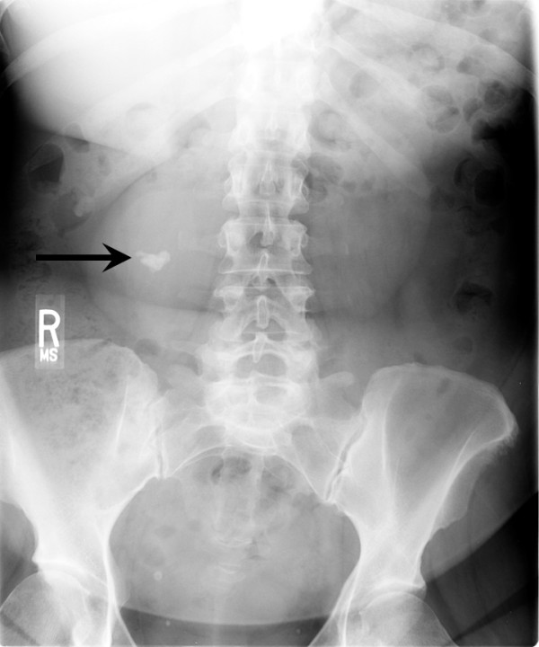
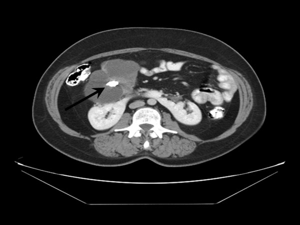
Methods: Here, we present a unique and first case of synchronous primary carcinoid tumor and moderately differentiated adenocarcinoma arising within mature cystic teratoma of horseshoe kidney in a 50-year-old female. Lumbar spine X-ray, done for her complaint of progressive chronic low back pain, accidentally found a large calcification overlying the lower pole of the right kidney. Further radiologic studies revealed horseshoe kidney and a large multi-septated cystic lesion immediately anterior to the right renal pelvis with central calcification and peripheral enhancement. She underwent right partial nephrectomy.

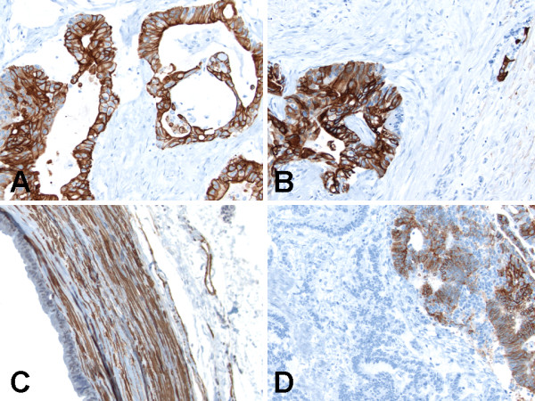
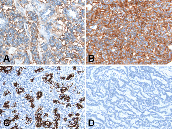
Results: Macroscopically, the encapsulated complex solid and multiloculated cystic tumor with large calcification, focal thickened walls and filled with yellow-tan gelatinous material. Microscopically, the tumor showed coexistent mature cystic teratoma, moderately differentiated adenocarcinoma and carcinoid tumor. Immunohistochemically, alpha-methylacyl-coenzyme A-racemase, calretinin, CD10 and thyroid transcription factor-1 were negative in all the three components of the tumor. The teratomatous cysts lined by ciliated epithelium showed strong staining for cytokeratin 7 and pancytokeratin, and those lined by colonic-like epithelium showed strong staining for CDX2, cytokeratin 20 and pancytokeratin, but both were negative for calretinin. Additionally, the teratomatous cyst wall showed strong staining for smooth muscle actin, and weak staining for carbonic anhydrase IX, CD99, chromogranin and synaptophysin. The adenocarcinoma component was strongly positive for cytokeratin 7 and pancytokeratin, weakly positive for synaptophysin and CD56, and negative for carbonic anhydrase IX, CD99, CDX2, chromogranin, cytokeratin 20 and smooth muscle actin. The carcinoid tumor component was strongly positive for CD56, chromogranin and synaptophysin, weakly positive for pancytokeratin, and negative for carbonic anhydrase IX, CD99, CDX2, cytokeratin 7, cytokeratin 20 and smooth muscle actin. She received no adjuvant therapy and is alive without evidence of disease six months after diagnosis and surgery.

Conclusion: This unique and first case herein presented with synchronous primary carcinoid tumor and primary adenocarcinoma arising within mature cystic teratoma of horseshoe kidney emphasizes the need for thorough sectioning and entire submission for histologic evaluation of mature cystic teratomas, in order to avoid missing multiple additional histogenetically distinct neoplasms.


Figures

Figure 1
Figure 1
Anterior-posterior view of lumbar X-ray showing a large calcification (arrow) overlying the lower pole of the right kidney. R indicates right side.
Figure 2
Figure 2
Computed tomography scan of abdomen and pelvis demonstrating horseshoe shaped kidney and a large multiseptated cystic lesion immediately anterior to the right renal pelvis with central calcification (arrow).
Figure 3
Figure 3
Histologic (hematoxylin and eosin stain) findings of synchronous primary carcinoid tumor and primary adenocarcinoma arising within mature cystic teratoma of horseshoe kidney. (A) Teratomatous component with multilocular cystic spaces. Original magnification ×20. (B) Teratomatous cyst lined by mucinous columnar enteric-type or colonic-like epithelium with muscular wall. Original magnification ×200. (C) Teratomatous cyst lined by ciliated epithelium with muscular wall. Original magnification ×600. (D) Teratomatous cyst wall composed predominantly of smooth muscle. Original magnification ×100.
Figure 4
Figure 4
Histologic (hematoxylin and eosin stain) findings of synchronous primary carcinoid tumor and primary adenocarcinoma arising within mature cystic teratoma of horseshoe kidney. (A) Adenocarcinoma component with infiltrating atypical glands. Original magnification ×200. (B) Adenocarcinoma component composed of cohesive pleomorphic epithelioid cells with adjacent necrosis. Original magnification ×100. (C) Adenocarcinoma component showing large cells with abundant eosinophilic cytoplasm, enlarged nuclei with contour irregularities, and occasional prominent nucleoli. Original magnification ×600. (D) Carcinoid tumor (left part of figure) and adenocarcinoma (right part of figure) components adjacent and closely apposed to each other with a clear transition zone. Original magnification ×100.
Figure 5
Figure 5
Histologic (hematoxylin and eosin stain) findings of synchronous primary carcinoid tumor and primary adenocarcinoma arising within mature cystic teratoma of horseshoe kidney. (A) Carcinoid tumor (left lower part of figure) and adenocarcinoma (right upper part of figure) components adjacent and closely apposed to each other without a clear transition zone. Original magnification ×100. (B) Carcinoid tumor showing the classical architectural pattern of trabecular nests of monotonous small round cells with peripheral palisading. Original magnification ×200. (C) Carcinoid tumor showing the classical architectural pattern of anastomosing ribbon-like nests of monotonous small round cells. Original magnification ×200. (D) Carcinoid tumor showing the classical cytologic features of fine granular "salt-and-pepper" chromatin pattern. Original magnification ×600.
Figure 6
Figure 6
Immunohistochemical (immunoperoxidase and hematoxylin counterstain) findings of synchronous primary carcinoid tumor and primary adenocarcinoma arising within mature cystic teratoma of horseshoe kidney. (A) CDX2 staining was positive in the mucinous columnar enteric-type or colonic-like epithelium lining of teratomatous cysts. Original magnification ×200. (B) Cytokeratin 20 staining was positive in the mucinous columnar enteric-type or colonic-like epithelium lining of teratomatous cysts. Original magnification ×200. (C) Smooth muscle actin was positive in teratomatous cyst wall. Original magnification ×200. (D) Cytokeratin 7 staining was positive in the adenocarcinoma component in right upper part of figure, but negative in the carcinoid tumor component in left lower part of figure. Original magnification ×200.
Figure 7
Figure 7
Immunohistochemical (immunoperoxidase and hematoxylin counterstain) findings of synchronous primary carcinoid tumor and primary adenocarcinoma arising within mature cystic teratoma of horseshoe kidney. (A) CD56 staining was positive in the carcinoid tumor component. Original magnification ×400. (B) Synaptophysin staining was positive in the carcinoid tumor component. Original magnification ×400. (C) Pancytokeratin staining was focally positive in the carcinoid tumor component. Original magnification ×200. (D) Cytokeratin 20 was negative in the carcinoid tumor component. Original magnification ×200.

References

    1. Rose PG, Tak WK, Reale FR. Squamous cell carcinoma arising in a mature cystic teratoma with metastasis to the paraaortic nodes. Gynecol Oncol. 1993;50:131–133. doi: 10.1006/gyno.1993.1178. - DOI - PubMed
    1. Griffiths D, Wass J, Look K, Sutton G. Malignant degeneration of a mature cystic teratoma five decades after discovery. Gynecol Oncol. 1995;59:427–429. doi: 10.1006/gyno.1995.9948. - DOI - PubMed
    1. Chadha S, Schaberg A. Malignant transformation in benign cystic teratomas: dermoids of the ovary. Eur J Obstet Gynecol Reprod Biol. 1988;29:329–338. doi: 10.1016/0028-2243(88)90074-3. - DOI - PubMed
    1. Resnick ME, Unterberger H, McLoughlin PT. Renal carcinoid producing the carcinoid syndrome. Med Times. 1966;94:895–896. - PubMed
    1. Raslan WF, Ro JY, Ordonez NG, Amin MB, Troncoso P, Sella A, Ayala AG. Primary carcinoid of the kidney. Immunohistochemical and ultrastructural studies of five patients. Cancer. 1993;72:2660–2666. doi: 10.1002/1097-0142(19931101)72:9<2660::AID-CNCR2820720923>3.0.CO;2-O. - DOI - PubMed