. 2009 Sep;35(3):352-8.
doi: 10.1016/j.nbd.2009.05.024. Epub 2009 Jun 10.

Soluble fibrillar oligomer levels are elevated in Alzheimer's disease brain and correlate with cognitive dysfunction

Affiliations

Soluble fibrillar oligomer levels are elevated in Alzheimer's disease brain and correlate with cognitive dysfunction

Jennifer L Tomic et al. Neurobiol Dis. 2009 Sep.

Abstract

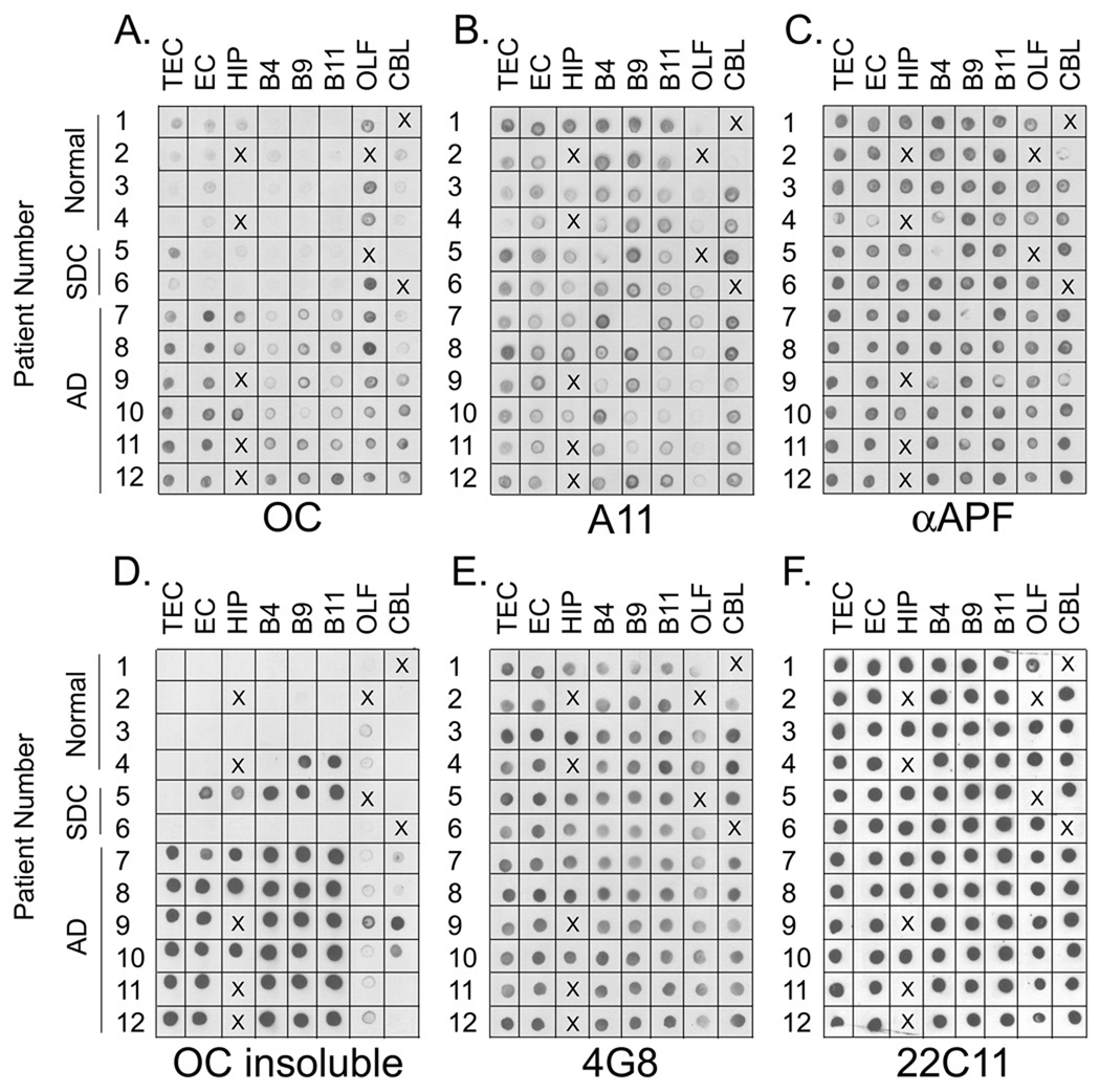
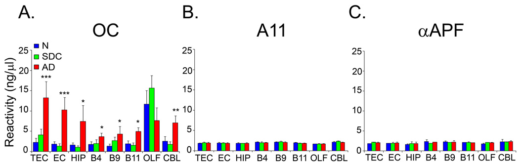
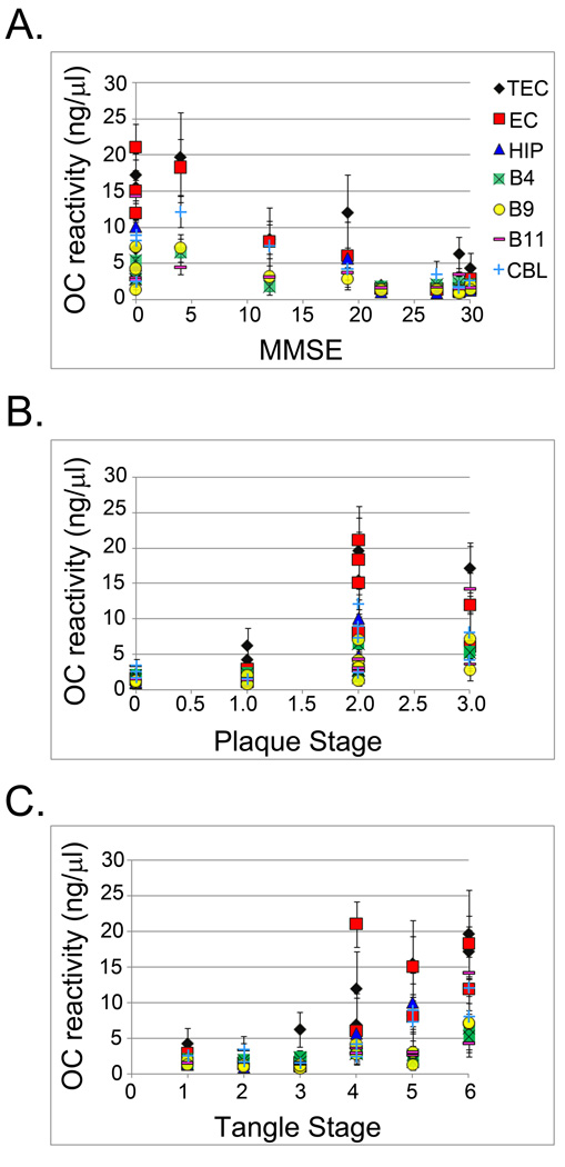
Recent evidence has suggested a role for soluble oligomeric Abeta species in the pathology of Alzheimer's disease (AD). Fibrillar plaque deposits are present in non-demented individuals and levels of soluble Abeta correlate better with cognitive dysfunction in AD and transgenic mouse models. We have previously reported that there are at least two conformationally distinct types of Abeta oligomers: prefibrillar oligomers that are kinetic intermediates in fibril assembly reactions and are specifically recognized by A11 antibody and fibrillar oligomers that may represent fibril seeds or small pieces of fibrils and are recognized by a fibril specific antibody, OC. We have examined the levels of these two types of oligomers in the PBS soluble fraction of brain tissue from control cases, cases with senile degenerative changes (SDC) and AD patients. We found that the levels of soluble fibrillar oligomers detected by OC antibody are significantly elevated in multiple brain regions of AD patients. The elevated fibrillar oligomer levels were found not to be an artifact of tissue homogenization, nor a result of increased Abeta or APP levels. The concentration of fibrillar oligomers in adjacent brain regions of the same patient can vary widely and were not detected in post-mortem cerebrospinal fluid. In contrast, the level of prefibrillar oligomers are variable in both AD and age matched controls, indicating that they are not correlated with cognitive dysfunction and suggesting that they precede dementia in AD. Significant correlations were found between the levels of fibrillar oligomers and cognitive decline (MMSE scores) as well as the neuropathological hallmarks of AD. These results indicate that fibrillar oligomers may play a key role in the pathology of AD and may be a new target for diagnostic and therapeutic development.


Figures

Figure 1
Figure 1. Soluble Oligomer levels in AD Brain
A. Dot blot analysis of PBS soluble fraction with the anti-fibril antibody ( OC). PBS soluble fractions from the transentorhinal cortex (TEC), entorhinal cortex (EC), hippocampus (HIP), Brodmann’s Area 4 (B4), Brodmann’s Area 9 (B9), Brodmann’s Area 11 (B11), olfactory bulb (OLF) and cerebellum (CBL) were spotted onto nitrocellulose membrane and reacted with the OC antibody. The most prominent immunoreactivity was found in all brain regions of AD cases while reduced or no immunoreactivity was present in normal controls or cases with senile degenerative changes (SDC). The signal present in these cases indicates the presence of soluble fibrillar oligomers. B, C. Dot blot analysis of prefibrillar oligomers with A11 antibody and annular protofibrils with αAPF antibody, respectively. Although prefibrillar oligomer levels are low in the olfactory bulb, no differences in the levels were observed in AD, SDC and normal controls. D. The 100,000 × G insoluble fractions were solublized with 8M urea, 50 mM Tris pH 7.4 and spotted onto nitrocellulose membrane for detection with OC antibody. Prominent immunoreactivity was found in all AD cases as well as two cases (4 and 5) with mild neuropathology. The presence of immunoreactivity in the insoluble fraction of these two cases and the lack of immunoreactivity in the PBS fractions of the same cases (A) indicates that the soluble fibrillar oligomers present in Figure 1a are not an artifact of homogenization. E, F. Dot blot analysis of PBS soluble fraction with 4G8 (anti-Aβ) and 22C11 (anti-APP) antibodies, respectively. The levels of total soluble Aβ do not differ significantly between AD, SDC and normal controls. The uniform distribution of 22C11 immunoreactivity indicates that equal amounts of lysate were spotted. X indicates that this sample was not available for that case.
Figure 2
Figure 2. Fibrillar oligomers are elevated in AD brain
Quantitative analysis of the dot blot data in Figure 1, showing human brain sample reactivity with A OC, B A11, and C. αAPF antibodies. A. Fibrillar oligomer levels were significantly higher in AD cases (AD, red bars) compared to normal (N, blue bars) and SDC (SDC, green bars) subjects in all the brain regions examined except OLF. B., C. No significant difference in A11 and αAPF reactivity between normal, SDC and AD samples were found. Human brain sample reactivity is expressed as ng/µl on the basis of a standard curve obtained using synthetic fibrillar oligomer, prefibrillar oligomer and annular protofibrillar preparations. Values are the mean ± SD of four independent experiments (* p ≤ 0.05; ** p ≤ 0.01, *** p≤ 0.001).
Figure 3
Figure 3. Soluble fibrillar oligomer levels in AD brain correlate with cognitive decline and neuropathology
A. OC immunoreactivity was compared to the MMSE, a measure of cognitive dysfunction. Significant inverse correlations were determined for TEC (black diamond), EC (red square), HIP (blue triangle), B4 (green square), B9 (yellow circle) and CBL (light blue cross). B. OC immunoreactivity was compared to the plaque stage and significant correlations between fibrillar oligomer levels and amyloid pathology were found in TEC, EC, HIP, B4 and B9. C. OC immunoreactivity was compared to the tangle stage and significant correlations between fibrillar oligomer levels and tau pathology were found in TEC, EC, HIP, B4, B9, B11 (purple dash) and CBL. The r and p values are reported in Table 2.

References

    1. Bancher C, Braak H, Fischer P, Jellinger KA. Neuropathological staging of Alzheimer lesions and intellectual status in Alzheime's and Parkinson’s disease patients. Neurosci Lett. 1993;162:179–182. - PubMed
    1. Barghorn S, Nimmrich V, Striebinger A, Krantz C, Keller P, Janson B, Bahr M, Schmidt M, Bitner RS, Harlan J, et al. Globular amyloid beta-peptide oligomer - a homogenous and stable neuropathological protein in Alzheime's disease. J Neurochem. 2005;95:834–847. - PubMed
    1. Bennett DA, Cochran EJ, Saper CB, Leverenz JB, Gilley DW, Wilson RS. Pathological changes in frontal cortex from biopsy to autopsy in Alzheime's disease. Neurobiol Aging. 1993;14:589–596. - PubMed
    1. Blessed G, Tomlinson BE, Roth M. The association between quantitative measures of dementia and of senile change in the cerebral grey matter of elderly subjects. Br J Psychiatry. 1968;114:797–811. - PubMed
    1. Braak H, Braak E. Neuropathological stageing of Alzheimer-related changes. Acta Neuropathol (Berl) 1991;82:239–259. - PubMed

Publication types

MeSH terms