. 2009 Jun 10;17(6):789-99.
doi: 10.1016/j.str.2009.04.006.

The P22 tail machine at subnanometer resolution reveals the architecture of an infection conduit

Affiliations

The P22 tail machine at subnanometer resolution reveals the architecture of an infection conduit

Gabriel C Lander et al. Structure. .

Abstract

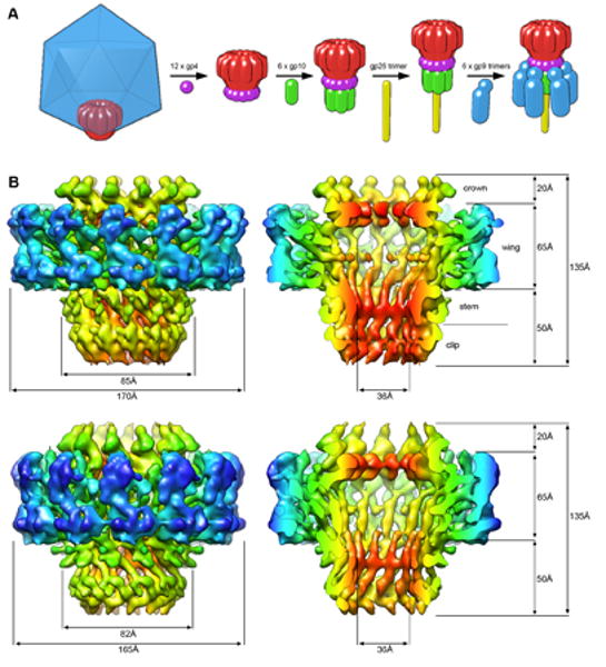
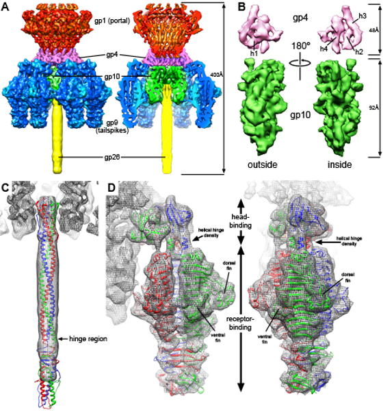
The portal channel is a key component in the life cycle of bacteriophages and herpesviruses. The bacteriophage P22 portal is a 1 megadalton dodecameric oligomer of gp1 that plays key roles in capsid assembly, DNA packaging, assembly of the infection machinery, and DNA ejection. The portal is the nucleation site for the assembly of 39 additional subunits generated from multiple copies of four gene products (gp4, gp10, gp9, and gp26), which together form the multifunctional tail machine. These components are organized with a combination of 12-fold (gp1, gp4), 6-fold (gp10, trimers of gp9), and 3-fold (gp26, gp9) symmetry. Here we present the 3-dimensional structures of the P22 assembly-naive portal formed from expressed subunits (gp1) and the intact tail machine purified from infectious virions. The assembly-naive portal structure exhibits a striking structural similarity to the structures of the portal proteins of SPP1 and phi29 derived from X-ray crystallography.


Figures

Figure 1
Figure 1
The tail machine assembly pathway and CryoEM structures of the 11-and 12-fold portals at sub nanometer resolution. A. The dodecameric portal (red) is shown at one pentameric vertex of the capsid (blue, not shown in subsequent steps). After DNA packaging, the tail assembles via sequential addition of multiple copies of gp4, gp10, gp26, and gp9 to the portal ring. B. Surface renderings of the reconstructed portal densities contoured at 1.5 sigma and colored radially from the central channel axis. The top two structures are of the 12-fold particles (8.6Å resolution), and the lower two of the 11-fold (8.8Å resolution). On the right are cutaway views of the portals, showing the arrangement of skewed helices in the stem region of the portals and a channel that is wide enough to accommodate double-stranded DNA. The same overall structural morphology is seen throughout both reconstructions.
Figure 2
Figure 2
Comparison of the P22 portal monomeric subunit with the SPP1 portal monomer, and three-dimensional mapping of the P22 portal sequence. A. Side views of the SPP1 portal (PDB entry: 2JES) are represented as ribbons colored from the N-terminus (blue) to C-terminus (red). B. Contoured at 1.5 sigma, the details of the P22 subunit extracted from the 12-fold portal structure shows many similarities to the subunit structure of the SPP1 portal. Both have crown, wing, stem, and clip regions, with P22 densities that are positioned similarly to helices 3, 4, and 6 of SPP1 (helical numbers defined in (Simpson et al, 2000)). The SPP1 subunit has been refined into the P22 density and is shown in the rightmost P22 portal density. P22 has an additional domain for which SPP1 has no counterpart (colored in magenta). C. Hydrogen-deuterium exchange data (Kang et al, 2008) has been combined with secondary structure prediction (www.predictprotein.org) and information from the SPP1 crystal structure to assign the location of the entire P22 sequence into the segmented P22 density. Domains are colored in the sequence according to their corresponding locations in the SPP1 ribbon representation, with the exception of the upper wing domain (magenta), which has no SPP1 counterpart. Residues after 580 were determined to be disordered and likely continue above the crown domain.
Figure 3
Figure 3
The P22 tail machine at 9.4Å resolution, as determined by cryoEM. A. Fifty-one subunits from five different gene products assemble to form the tail machine, and are represented in surface renderings at 1.5 sigma. The image on the right depicts a cutaway view to expose the tail machine interior. The gene products are colored as follows: gp1 are red, gp4 are magenta, gp10 are green, gp9 are blue, gp26 are yellow. B. The exterior and interior views of the segmented gp4 (above, magenta) and gp10 (below, green) monomeric densities. The gp4 density, which is predicted to consist of four alpha helices, exhibits four distinctly sausage-like densities that are characteristic of alpha helices. Two of these run laterally at the gp10 interface (h1 and h2), and the other two run parallel to the channel axis on the interior of the monomer (h3 and h4). These two interior helices appear to interact with gp1 and gp10 in the reconstructed density. The gp10 density is much more difficult to interpret that the gp4 density, and secondary structure prediction shows that this protein is mostly made up of beta strands. C. The gp26 tail needle (PDB entry: 2POH) is docked into the EM density, showing extensive interactions with gp10 at the N-terminal tip. Further from the tail machine body, the needle density becomes more disordered, becoming completely disordered shortly after passing the hinge domain. D. The crystal structures for the N-terminal head-binding domain and the C-terminal receptor-binding domain of gp9 (PDB entries: 1LKT, 1TYU) are docked into the EM density, fitting with high fidelity. The tail spike assumes an asymmetric organization upon binding to the tail machine with the head-binding domain tilted relative to the axis of the tail machine. Two of the head-binding subunits are involved in interactions that bridge gp4 and gp10, while the third subunit interacts with the three N-terminal helices of the receptor-binding domain.

References

    1. Ackermann HW. Tailed bacteriophages: the order caudovirales. Adv Virus Res. 1998;51:135–201. - PMC - PubMed
    1. Agirrezabala X, Martin-Benito J, Caston JR, Miranda R, Valpuesta JM, Carrascosa JL. Maturation of phage T7 involves structural modification of both shell and inner core components. EMBO J. 2005a;24(21):3820–3829. - PMC - PubMed
    1. Agirrezabala X, Martin-Benito J, Valle M, Gonzalez JM, Valencia A, Valpuesta JM, Carrascosa JL. Structure of the connector of bacteriophage T7 at 8A resolution: structural homologies of a basic component of a DNA translocating machinery. Journal of molecular biology. 2005b;347(5):895–902. - PubMed
    1. Bazinet C, Benbasat J, King J, Carazo JM, Carrascosa JL. Purification and organization of the gene 1 portal protein required for phage P22 DNA packaging. Biochemistry. 1988;27(6):1849–1856. - PubMed
    1. Bhardwaj A, Olia AS, Walker-Kopp N, Cingolani G. Domain organization and polarity of tail needle GP26 in the portal vertex structure of bacteriophage P22. Journal of molecular biology. 2007;371(2):374–387. - PubMed

Publication types

MeSH terms